File:Micrograph of bullous pemphigoid.jpg
Jump to navigation
Jump to search

Size of this preview: 800 × 593 pixels. Other resolutions: 320 × 237 pixels | 640 × 474 pixels | 1,024 × 759 pixels | 1,280 × 948 pixels | 1,401 × 1,038 pixels.
Original file (1,401 × 1,038 pixels, file size: 342 KB, MIME type: image/jpeg)
Summary
| Description |
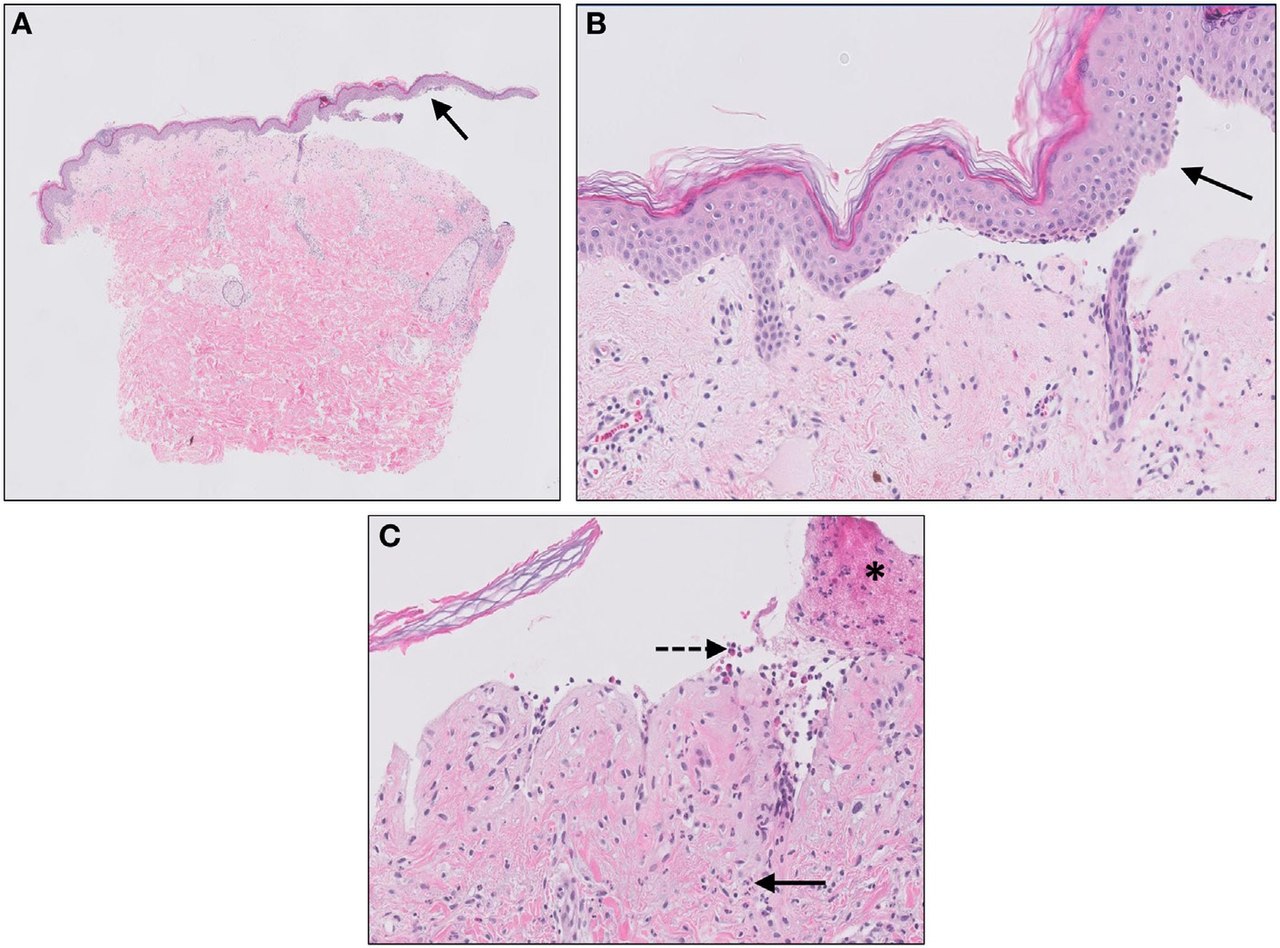
English: Micrograph of bullous pemphigoid. Subepidermal blistering [solid arrows in (A,B)] and influx of inflammatory cells including eosinophils and neutrophils in the dermis [solid arrow (C)] and blister cavity [dashed arrows (C)]. In (C) also deposition of fibrin is noted (asterisks). |
| Date | |
| Source |
(2018). "Complement Activation in Inflammatory Skin Diseases". Frontiers in Immunology 9. DOI:10.3389/fimmu.2018.00639. ISSN 1664-3224.
|
| Author | Jenny Giang, Marc A. J. Seelen, Martijn B. A. van Doorn, Robert Rissmann,Errol P. Prens and Jeffrey Damman |
Licensing
This file is licensed under the Creative Commons Attribution 4.0 International license.
- You are free:
- to share – to copy, distribute and transmit the work
- to remix – to adapt the work
- Under the following conditions:
- attribution – You must give appropriate credit, provide a link to the license, and indicate if changes were made. You may do so in any reasonable manner, but not in any way that suggests the licensor endorses you or your use.
File history
Click on a date/time to view the file as it appeared at that time.
| Date/Time | Thumbnail | Dimensions | User | Comment | |
|---|---|---|---|---|---|
| current | 12:28, 5 November 2019 | | 1,401 × 1,038 (342 KB) | commons>Mikael Häggström | User created page with UploadWizard |
File usage
The following page uses this file: