File:Juxta-cortical-chondroma.png
Juxta-cortical-chondroma.png (333 × 560 pixels, file size: 164 KB, MIME type: image/png)
Summary
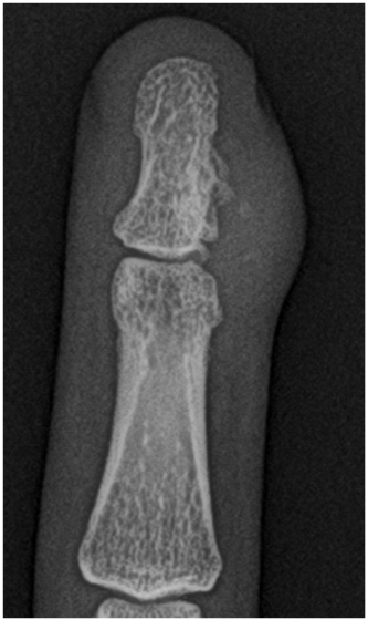
Author: Case courtesy of Dr Dalia Ibrahim, Radiopaedia.org, rID: 27609 Source: https://radiopaedia.org/cases/juxta-cortical-chondroma?lang=gb Description: X-ray finger- Right middle finger distal phalangeal swelling (age 38). Well defined soft tissue mass showing foci of calcfications and adjacent bony remodelling, it erodes and induces sclerosis in the contiguous cortical bone producing the characterstic "Pop corn" apperance.Diagnosis confirmed at biopsy.
Licensing
| This work is licensed under the Creative Commons Attribution-NonCommersial-ShareAlike 4.0 License. |
File history
Click on a date/time to view the file as it appeared at that time.
| Date/Time | Thumbnail | Dimensions | User | Comment | |
|---|---|---|---|---|---|
| current | 05:27, 15 June 2021 |  | 333 × 560 (164 KB) | Whispyhistory (talk | contribs) | Author: Case courtesy of Dr Dalia Ibrahim, Radiopaedia.org, rID: 27609 Source: https://radiopaedia.org/cases/juxta-cortical-chondroma?lang=gb Description: X-ray finger- Right middle finger distal phalangeal swelling (age 38). Well defined soft tissue mass showing foci of calcfications and adjacent bony remodelling, it erodes and induces sclerosis in the contiguous cortical bone producing the characterstic "Pop corn" apperance.Diagnosis confirmed at biopsy. |
You cannot overwrite this file.
File usage
The following file is a duplicate of this file (more details):
- File:Juxta-cortical chondroma (Radiopaedia 27609-27821 Frontal Zoomed in 1).png from a shared repository
There are no pages that use this file.
