File:Histopathology of epidermoid cyst.jpg
Jump to navigation
Jump to search

Size of this preview: 799 × 322 pixels. Other resolutions: 320 × 129 pixels | 1,027 × 414 pixels.
Original file (1,027 × 414 pixels, file size: 138 KB, MIME type: image/jpeg)
Summary
| Description |
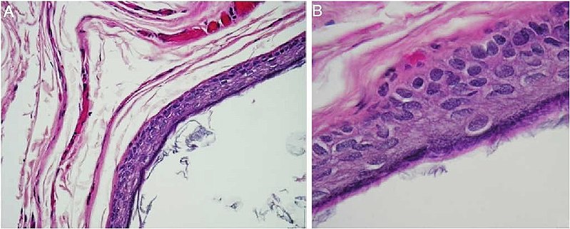
English: Histopathology of epidermoid cyst: (A) Cyst wall lined by keratinizing stratified squamous epithelium and contains keratin flakes (Hematoxylin and Eosin stain, magnification 100×); (B) Cyst wall lined by keratinizing stratified squamous epithelium (Hematoxylin and Eosin stain, magnification 400×).. From the same case:
|
| Date | |
| Source |
(2020). "Epidermoid cyst of the suprasternal region: a rare case report". Brazilian Journal of Otorhinolaryngology 86 (1): 133–135. DOI:10.1016/j.bjorl.2016.04.010. ISSN 18088694. Attribution 4.0 International (CC BY 4.0) license |
| Author | Bin Manie, Manal Al; Al-Qahtani, Khalid Hussain; Al Ammar, Ahmed; Islam, Tahera; Otaibi, Faiza N. Al |


Licensing
This file is licensed under the Creative Commons Attribution 4.0 International license.
- You are free:
- to share – to copy, distribute and transmit the work
- to remix – to adapt the work
- Under the following conditions:
- attribution – You must give appropriate credit, provide a link to the license, and indicate if changes were made. You may do so in any reasonable manner, but not in any way that suggests the licensor endorses you or your use.
File history
Click on a date/time to view the file as it appeared at that time.
| Date/Time | Thumbnail | Dimensions | User | Comment | |
|---|---|---|---|---|---|
| current | 12:37, 4 March 2020 | 1,027 × 414 (138 KB) | commons>Mikael Häggström | User created page with UploadWizard |
File usage
There are no pages that use this file.