File:Gr4 lrgnih.jpg

From WikiProjectMed
Jump to navigation Jump to search

Gr4_lrgnih.jpg(793 × 591 pixels, file size: 89 KB, MIME type: image/jpeg)

This file is from a shared repository and may be used by other projects. The description on its file description page there is shown below.

Summary

Description
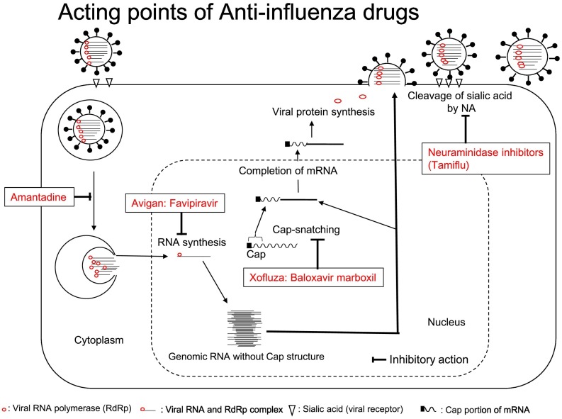
English⧼Colon⧽ Replication cycle of influenza and sites of action of anti-influenza drugs. Influenza virus hemagglutinin (HA) binds to its receptor, sialic acid on the cell surface and is taken up into endosomes by endocytosis. Endosomes are gradually acidified to produce late endosomes, viral matrix-2 (M2) ion channels acidify viral particles, and the structure of trimeric HA molecules changes and shows membrane fusion activity. The endosomal membrane and envelope are then fused, and the genomic RNA and RdRp complex in the virus particle are released into the cytoplasm (uncoating) and transported to the nucleus through the cytoplasm. The transcription (replication) of genomic RNA by the RdRp complex occurs in the nucleus, and the genomic RNA is abundantly produced. Genomic RNA lacks the Cap structure required for mRNA function, and the Cap portion of the host mRNA is removed by a Cap-dependent endonuclease. Then, the Cap portion is coupled to genomic RNA, and viral mRNA synthesis is complete (Cap-snatching). Viral proteins are synthesized from the mRNA, and viral proteins and genomic RNA are transported to the cell surface and bud from the membrane to form viral particles. Since the budding virus is bound to the sialic acid on the infected cell surface via the HA protein of virus particles, it is unable to leave the infected cell and infect new cells. For this reason, the sialic acid-HA bond is cleaved by neuraminidase (NA) on the surface of the virus particle, and the virus particle is released from the surface of the infected cell and proceeds to the next round of infection. Amantadine blocks uncoating by inhibiting acidification mediated by the M2 protein in the virus particle in late endosome, and thus the infection is unable to be completed. Favipiravir inhibits viral RNA synthesis by terminating chain elongation at its incorporated site, and no new RNA is generated in the cell. Baloxavir marboxil prevents Cap-snatching, and the viral mRNA is not produced, resulting in a failure to produce viral proteins and infectious viruses. Genomic RNA is synthesized and remains in the cell. NA inhibitors block the cleavage of the sialic acid-HA bond in the virus on the surface of infected cells and prevent the spread of the viral infection. NA inhibitors allow genomic RNA synthesis. Favipiravir inhibits viral RNA synthesis, while baloxavir and NAIs allow viral RNA synthesis. Although viral spread is inhibited by baloxavir and NAIs, viral RNA is synthesized, and the pool of genomic RNA serves as a rich source of resistant viruses.
Date
Source https://www.ncbi.nlm.nih.gov/pmc/articles/PMC7102570/
Author Kimiyasu Shirakia, Tohru Daikokub

Licensing

English⧼Colon⧽ This file is licensed CC BY-NC 3.0

This file was uploaded with UploadWizard.

File history

Click on a date/time to view the file as it appeared at that time.

Date/TimeThumbnailDimensionsUserComment
current16:46, 20 May 2023Thumbnail for version as of 16:46, 20 May 2023793 × 591 (89 KB)Ozzie10aaaaUploaded a work by Kimiyasu Shirakia, Tohru Daikokub from https://www.ncbi.nlm.nih.gov/pmc/articles/PMC7102570/ with UploadWizard

There are no pages that use this file.
Metadata