File:Fibrous-dysplasia-femur-and-pelvis.jpg
Fibrous-dysplasia-femur-and-pelvis.jpg (655 × 512 pixels, file size: 69 KB, MIME type: image/jpeg)
Summary
Author: Case courtesy of Dr Michael P Hartung, Radiopaedia.org, rID: 74812
Source: https://radiopaedia.org/cases/fibrous-dysplasia-femur-and-pelvis?lang=gb
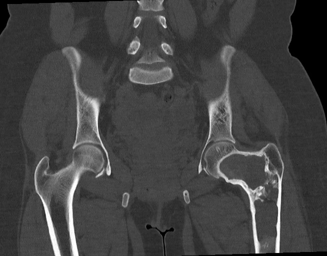
Description: CT scan showing 25 year old female with left hip pain. Shepherd crook deformity of the left femur with a large, irregular lucent lesion expanding the femoral neck through proximal diaphysis. Some early cortical breakthrough along the anterior margin. Few smaller lucencies in the left pelvis/acetabulum.
Licensing
| This work is licensed under the Creative Commons Attribution-NonCommersial-ShareAlike 4.0 License. |
File history
Click on a date/time to view the file as it appeared at that time.
| Date/Time | Thumbnail | Dimensions | User | Comment | |
|---|---|---|---|---|---|
| current | 13:31, 14 May 2021 | | 655 × 512 (69 KB) | Whispyhistory (talk | contribs) | Author: Case courtesy of Dr Michael P Hartung, Radiopaedia.org, rID: 74812 Source: https://radiopaedia.org/cases/fibrous-dysplasia-femur-and-pelvis?lang=gb Description: CT scan showing 25 year old female with left hip pain. Shepherd crook deformity of the left femur with a large, irregular lucent lesion expanding the femoral neck through proximal diaphysis. Some early cortical breakthrough along the anterior margin. Few smaller lucencies in the left pelvis/acetabulum. |
You cannot overwrite this file.
File usage
The following file is a duplicate of this file (more details):
- File:Fibrous dysplasia - femur and pelvis (Radiopaedia 74812-85821 Coronal 1).jpg from a shared repository
.jpg)
There are no pages that use this file.