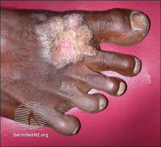
File:DermNet NZ chromoblastomycosis-04.png

From WikiProjectMed
Jump to navigation Jump to search

DermNet_NZ_chromoblastomycosis-04.png(516 × 471 pixels, file size: 444 KB, MIME type: image/png)

This file is from a shared repository and may be used by other projects. The description on its file description page there is shown below.

Summary

Description: Histopathologyof chromoblastomycosismay show typical thick-walled dark-brown 'sclerotic' cells on skinbiopsyconfirming the presence of a dematiaceous fungus. It is dark coloured due tomelaninin the walls of the organism.
Source: dermnetnz.org
Gallery page: https://dermnetnz.org/topics/chromoblastomycosis
Author: DermNet New Zealand
Date: 2021-02-18

License

Attribution-NonCommercial-NoDerivs 3.0 New Zealand (CC BY-NC-ND 3.0 NZ)

File history

Click on a date/time to view the file as it appeared at that time.

Date/TimeThumbnailDimensionsUserComment
current01:46, 21 April 2021Thumbnail for version as of 01:46, 21 April 2021516 × 471 (444 KB)Transfer to NC Commons from mdwiki

There are no pages that use this file.
Metadata