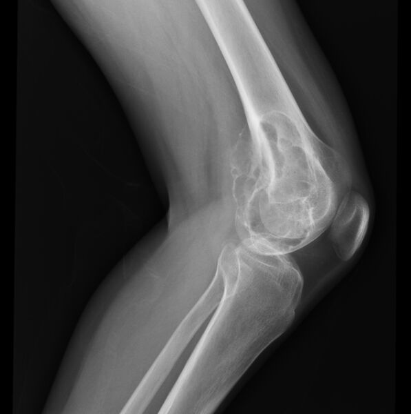
File:Chondromyxoid-fibroma-2.jpg

Original file (2,028 × 2,037 pixels, file size: 310 KB, MIME type: image/jpeg)
Summary
Author:Case courtesy of Dr Yasser Asiri, Radiopaedia.org, rID: 64932 Source:https://radiopaedia.org/cases/chondromyxoid-fibroma-2?lang=gb Description:X-ray knee- An eccentric bubbly and lucent lesion in the distal femoral metaphysis and epiphysis with sclerotic margins and narrow zone of transition in close proximity to the articular surface. There is ill-definition of the cortex in the medial aspect of the lesion, suspicious for cortical breakthrough. No suspicious periosteal reaction or pathological fracture.
Licensing
| This work is licensed under the Creative Commons Attribution-NonCommersial-ShareAlike 4.0 License. |
File history
Click on a date/time to view the file as it appeared at that time.
| Date/Time | Thumbnail | Dimensions | User | Comment | |
|---|---|---|---|---|---|
| current | 12:51, 5 July 2021 |  | 2,028 × 2,037 (310 KB) | Whispyhistory (talk | contribs) | Author:Case courtesy of Dr Yasser Asiri, Radiopaedia.org, rID: 64932 Source:https://radiopaedia.org/cases/chondromyxoid-fibroma-2?lang=gb Description:X-ray knee- An eccentric bubbly and lucent lesion in the distal femoral metaphysis and epiphysis with sclerotic margins and narrow zone of transition in close proximity to the articular surface. There is ill-definition of the cortex in the medial aspect of the lesion, suspicious for cortical breakthrough. No suspicious periosteal reaction or pat... |
You cannot overwrite this file.
File usage
The following file is a duplicate of this file (more details):
- File:Chondromyxoid fibroma (Radiopaedia 64932-73885 Lateral 1).jpg from a shared repository
The following 2 pages use this file: