File:Chondroblastoma-distal-femur-2-2.jpg

Original file (1,024 × 1,024 pixels, file size: 89 KB, MIME type: image/jpeg)
Summary
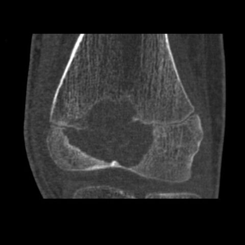
Author: Case courtesy of Dr Angela Byrne, Radiopaedia.org, rID: 8112
Source: https://radiopaedia.org/cases/chondroblastoma-distal-femur-2?lang=us
Description: CT confirms the plain film appearances, revealing a sharply demarcated epiphyseal lucent lesion but with faintly sclerotic margins. It transgresses the growth plate into the anterior part of the metaphysis. There is no periosteal reaction, however the does appear to be a cortical breach anterosuperiorly into the knee joint. No matrix calcification or extra-osseous soft tissue component can be demonstrated.
Licensing
| This work is licensed under the Creative Commons Attribution-NonCommersial-ShareAlike 4.0 License. |
File history
Click on a date/time to view the file as it appeared at that time.
| Date/Time | Thumbnail | Dimensions | User | Comment | |
|---|---|---|---|---|---|
| current | 12:32, 9 May 2021 | | 1,024 × 1,024 (89 KB) | Whispyhistory (talk | contribs) | Author: Case courtesy of Dr Angela Byrne, Radiopaedia.org, rID: 8112 Source: https://radiopaedia.org/cases/chondroblastoma-distal-femur-2?lang=us Description: CT confirms the plain film appearances, revealing a sharply demarcated epiphyseal lucent lesion but with faintly sclerotic margins. It transgresses the growth plate into the anterior part of the metaphysis. There is no periosteal reaction, however the does appear to be a cortical breach anterosuperiorly into the knee joint. No matrix... |
You cannot overwrite this file.
File usage
The following file is a duplicate of this file (more details):
- File:Chondroblastoma - distal femur (Radiopaedia 8112-8950 Coronal bone window 1).jpg from a shared repository
.jpg)
There are no pages that use this file.