File:COVID19CT1.webp
Jump to navigation
Jump to search

Size of this PNG preview of this WEBP file: 800 × 282 pixels. Other resolutions: 320 × 113 pixels | 1,179 × 415 pixels.
Original file (1,179 × 415 pixels, file size: 127 KB, MIME type: image/webp)
Summary
| Description |
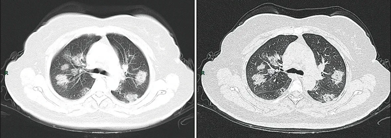
English: CT imaging of rapid progression stage. A 50 years old female with anorexia, fatigue, muscle soreness, nasal congestion and runny nose for 1 week, sore and itching throat for 2 days. Laboratory test: increased erythrocyte sedimentation rate (25 mm/h), normal white blood cells (4.08 × 10⁹/L), decreased lymphocytes (0.96 × 10⁹/ L), increased C-reaction protein (60.8 mg/L). Imaging examination: a (thin layer CT) and b (high-resolution CT) showed multiple patchy and light consolidation in both lungs and grid-like thickness of interlobular septa
Русский: КТ-снимок при быстропрогрессирующей болезни. 50-летняя женщина с анорексией, слабостью, ломотой в мышцах,заложенностью носа и насморком в течение 1 недели, болью и першением в горле в течение 2 дней. Лабораторный тест: повышенная скорость оседания эритроцитов (25 мм/ч), нормальный уровень лейкоцитов (4.08 × 10⁹/л), пониженный уровень лимфоцитов (0.96 × 10⁹/ л), повышенное количество С-реактивного белка (60.8 мг/л). Снимки: а (тонкослойная КТ) и b (КТ с высоким разрешением) показывают множественную фрагментарную и лёгкую консолидацию в обоих лёгких и сетчатую толщину междольковых перегородок |
| Date | |
| Source | https://mmrjournal.biomedcentral.com/articles/10.1186/s40779-020-0233-6 |
| Author | Jin, Y., Cai, L., Cheng, Z. et al. |
Licensing
This file is licensed under the Creative Commons Attribution 4.0 International license.
- You are free:
- to share – to copy, distribute and transmit the work
- to remix – to adapt the work
- Under the following conditions:
- attribution – You must give appropriate credit, provide a link to the license, and indicate if changes were made. You may do so in any reasonable manner, but not in any way that suggests the licensor endorses you or your use.
File history
Click on a date/time to view the file as it appeared at that time.
| Date/Time | Thumbnail | Dimensions | User | Comment | |
|---|---|---|---|---|---|
| current | 05:58, 9 March 2020 | 1,179 × 415 (127 KB) | commons>Jmarchn | Crop image |
File usage
The following page uses this file: