File:Axenfeld syndrome.jpg
Jump to navigation
Jump to search
Size of this preview: 504 × 599 pixels. Other resolutions: 202 × 240 pixels | 404 × 480 pixels | 646 × 768 pixels | 1,200 × 1,426 pixels.
Original file (1,200 × 1,426 pixels, file size: 396 KB, MIME type: image/jpeg)
| Description |
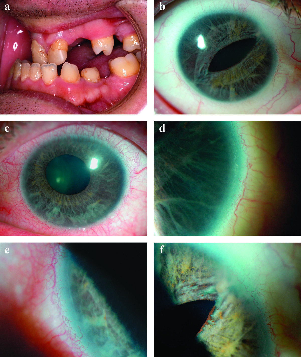
English: 1a Microdontia and hypodontia. 1b Slit pupil and iris atrophy right eye. 1c Corectopia with iris atrophy left eye. 1d Posterior embryotoxon right eye. 1e Posterior embryotoxon left eye. 1f Broad peripheral anterior synechiae right eye. |
| Date | |
| Source | L Dhir, K Frimpong-Ansah, Nabil E Habib: Missed case of Axenfeld-Rieger syndrome: a case report. Cases Journal 2008, 1:299 (6 November 2008) |
| Author | see below |
| Permission (Reusing this file) |
[1] |
This file is licensed under the Creative Commons Attribution 2.0 Generic license.
- You are free:
- to share – to copy, distribute and transmit the work
- to remix – to adapt the work
- Under the following conditions:
- attribution – You must give appropriate credit, provide a link to the license, and indicate if changes were made. You may do so in any reasonable manner, but not in any way that suggests the licensor endorses you or your use.
File history
Click on a date/time to view the file as it appeared at that time.
| Date/Time | Thumbnail | Dimensions | User | Comment | |
|---|---|---|---|---|---|
| current | 17:56, 13 January 2009 | | 1,200 × 1,426 (396 KB) | commons>Filip em | {{Information |Description={{en|1=1a Microdontia and hypodontia. 1b Slit pupil and iris atrophy right eye. 1c Corectopia with iris atrophy left eye. 1d Posterior embryotoxon right eye. 1e Posterior embryotoxon left eye. 1f Broad peripheral anterior synech |
File usage
There are no pages that use this file.