File:Animal Cell.svg

From WikiProjectMed
Jump to navigation Jump to search

Original file(SVG file, nominally 1,405 × 811 pixels, file size: 457 KB)

This file is from a shared repository and may be used by other projects. The description on its file description page there is shown below.

Summary

Description
English: A reworked version of File:Biological_cell.svg.

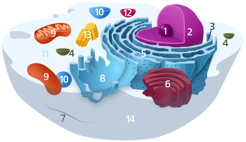
Diagram of a typical animal cell. Organelles are labelled as follows:

  1. Nucleolus
  2. Nucleus
  3. Ribosomes (dots on rough reticulum walls)
  4. Vesicle
  5. Rough endoplasmic reticulum
  6. Golgi apparatus (or "Golgi body")
  7. Cytoskeleton
  8. Smooth endoplasmic reticulum
  9. Mitochondrion
  10. Vacuole
  11. Cytosol
  12. Lysosome
  13. Centriole
  14. Cell membrane
Svenska: Schematisk bild över en typisk eukaryot cell, som visar cellens subcellulära komponenter. Organeller:
  1. Nukleol
  2. Cellkärna
  3. Ribosom
  4. Vesikel
  5. Korniga endoplasmatiska nätverket
  6. Golgiapparaten
  7. Cellskelettet
  8. Glatta endoplasmatiska nätverket
  9. Mitokondrie
  10. Vakuol
  11. Cytoplasma
  12. Lysosom
  13. Centriol
  14. Cellmembran
Deutsch: Organisation einer typischen eukaryotischen Tierzelle:
  1. de:Nucleolus
  2. de:Nukleus
  3. de:Ribosomen
  4. de:Vesikel
  5. Raues (Granuläres) ER (Ergastoplasma)
  6. Golgi-Apparat
  7. Cytoskelett
  8. Glattes (Agranuläres) ER
  9. Mitochondrien
  10. Lysosom
  11. Cytoplasma (mit Cytosol und Cytoskelett)
  12. Peroxisomen
  13. Zentriolen
  14. Zellmembran
Date
Source Own work
Author Kelvinsong
Permission
(Reusing this file)
I, the copyright holder of this work, hereby publish it under the following license:
Creative Commons CC-Zero This file is made available under the Creative Commons CC0 1.0 Universal Public Domain Dedication.
The person who associated a work with this deed has dedicated the work to the public domain by waiving all of their rights to the work worldwide under copyright law, including all related and neighboring rights, to the extent allowed by law. You can copy, modify, distribute and perform the work, even for commercial purposes, all without asking permission.

Captions

Components of a typical animal cell

Items portrayed in this file

depicts

1 December 2012

image/svg+xml

437a1e698973ab23dc94b4e6087e42a17b44f38a

468,171 byte

811 pixel

1,405 pixel

File history

Click on a date/time to view the file as it appeared at that time.

Date/TimeThumbnailDimensionsUserComment
current14:47, 17 November 2022Thumbnail for version as of 14:47, 17 November 20221,405 × 811 (457 KB)commons>TheBartgryReverted to version as of 00:21, 10 December 2012 (UTC) showing continuity between nuclear membrane and ER is useful

There are no pages that use this file.

Metadata