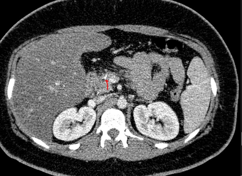
File:Acute on chronic pancreatitis (Radiopaedia 80902-94538 B 1).JPG

From WikiProjectMed
Jump to navigation Jump to search

Original file(814 × 594 pixels, file size: 103 KB, MIME type: image/jpeg)

This file is from a shared repository and may be used by other projects. The description on its file description page there is shown below.

Summary:

Description
  • Radiopaedia case ID: 80902
  • Image ID: 53278412
  • Plane projection: axial portal phase
  • Study findings: The blue and black arrows point to dilated main pancreatic duct and peripancreatic minimal fat stranding, respectively. The red arrow depicts the stones.
  • Modality: Annotated image
  • System: Hepatobiliary
  • Findings: Evidence of previous cholecystectomy and hepatosteatosis is seen. Mild peripancreatic fat stranding is noted in favor of acute pancreatitis. Three calcified stones are visible within the pancreatic duct at the neck with dilation of the distal main pancreatic duct suggestive of chronic pancreatitis. The proximal pancreatic duct in the head appears normal. A small hypo-enhancing area in the pancreatic tail may represent necrosis. No fluid collection or biliary dilation depicted.
Date Published: 18th Aug 2020
Source https://radiopaedia.org/cases/acute-on-chronic-pancreatitis-2
Author Faeze Salahshour
Permission
(Permission-reusing-text)
http://creativecommons.org/licenses/by-nc-sa/3.0/

Licensing:

Attribution-NonCommercial-ShareAlike 3.0 Unported (CC BY-NC-SA 3.0)

File history

Click on a date/time to view the file as it appeared at that time.

Date/TimeThumbnailDimensionsUserComment
current21:39, 15 April 2021Thumbnail for version as of 21:39, 15 April 2021814 × 594 (103 KB)Radiopaedia project rID:80902 (batch #955-2 B1)

The following page uses this file:

Metadata