File:5057022555 cabcf6d00a c.jpg

From WikiProjectMed
Jump to navigation Jump to search

Original file(647 × 800 pixels, file size: 163 KB, MIME type: image/jpeg)

This file is from a shared repository and may be used by other projects. The description on its file description page there is shown below.

Summary

Description
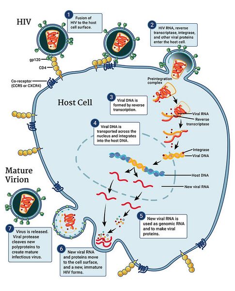
English⧼Colon⧽ This infographic illustrates the HIV replication cycle, which begins when HIV fuses with the surface of the host cell. A capsid containing the virus’s genome and proteins then enters the cell. The shell of the capsid disintegrates and the HIV protein called reverse transcriptase transcribes the viral RNA into DNA. The viral DNA is transported across the nucleus, where the HIV protein integrase integrates the HIV DNA into the host’s DNA. The host’s normal transcription machinery transcribes HIV DNA into multiple copies of new HIV RNA. Some of this RNA becomes the genome of a new virus, while the cell uses other copies of the RNA to make new HIV proteins. The new viral RNA and HIV proteins move to the surface of the cell, where a new, immature HIV forms. Finally, the virus is released from the cell, and the HIV protein called protease cleaves newly synthesized polyproteins to create a mature infectious virus. Credit: NIAID
Date
Source https://www.flickr.com/photos/niaid/5057022555/
Author NIAID

Licensing

{{subst:Custom license marker added by UW}} https://creativecommons.org/licenses/by/2.0/ Attribution 2.0 Generic (CC BY 2.0)

This file was uploaded with UploadWizard.

File history

Click on a date/time to view the file as it appeared at that time.

Date/TimeThumbnailDimensionsUserComment
current22:33, 26 March 2023Thumbnail for version as of 22:33, 26 March 2023647 × 800 (163 KB)Ozzie10aaaaUploaded a work by NIAID from https://www.flickr.com/photos/niaid/5057022555/ with UploadWizard

The following page uses this file:

Metadata