Fibrosarcoma
| Fibrosarcoma | |
|---|---|
 | |
| Micrograph of a tumour with the herringbone pattern as may be seen in fibrosarcoma. H&E stain. | |
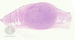
Fibrosarcoma (fibroblastic sarcoma) is a malignant mesenchymal tumour derived from fibrous connective tissue and characterized by the presence of immature proliferating fibroblasts or undifferentiated anaplastic spindle cells in a storiform pattern. In humans it is usually found in males aged 30 to 40.[citation needed] It originates in fibrous tissues of the bone and invades long or flat bones such as the femur, tibia, and mandible. It also involves the periosteum and overlying muscle.
Symptoms and signs
-
pathology-Fibrosarcoma
-
pathology-Fibrosarcoma
-
pathology-Fibrosarcoma
-
pathology-Fibrosarcoma
.jpg)
.jpg)
.jpg)
.jpg)
Adult-type
Individuals presenting with fibrosarcoma are usually adults thirty to fifty-five years old, often presenting with pain. Among adults, males have a higher incidence for fibrosarcoma than females.[citation needed]
Infantile-type
In infants, fibrosarcoma (often termed congenital infantile fibrosarcoma) is usually congenital. Infants presenting with this fibrosarcoma usually do so in the first two years of their life. Cytogenetically, congenital infantile fibrosarcoma is characterized by the majority of cases having a translocation between chromosomes 12 and 15 (notated as t(12;15)(p13;q25)) that results in formation of the fusion gene, ETV6-NTRK3, plus individual cases exhibiting trisomy for chromosomes 8, 11, 17, or 20.[1] The histology, association with the ETV6-NRTK3 fusion gene as well as certain chromosome trisomies, and the distribution of markers for cell type (i.e. cyclin D1 and Beta-catenin) within this tumor are similar to those found in the cellular form of mesoblastic nephroma. Indeed, mesoblastic nephroma and congenital infantile sarcoma appear to be the same disease with the exception that mesoblastic lymphoma originates in the kidney whereas congenital infantile sarcoma originates in non-renal tissues.[2][3][4]
Pathology
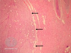
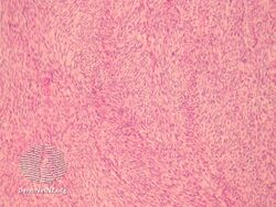
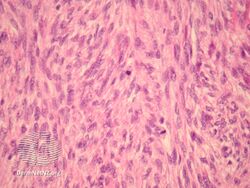
The tumor may present different degrees of differentiation: low grade (differentiated), intermediate malignancy and high malignancy (anaplastic). Depending on this differentiation, tumour cells may resemble mature fibroblasts (spindle-shaped), secreting collagen, with rare mitoses. These cells are arranged in short fascicles which split and merge, giving the appearance of "fish bone" known as a herringbone pattern. Poorly differentiated tumors consist in more atypical cells, pleomorphic, giant cells, multinucleated, numerous atypical mitoses and reduced collagen production. Presence of immature blood vessels (sarcomatous vessels lacking endothelial cells) favors the bloodstream metastasizing. There are many tumors in the differential diagnosis, including spindle cell melanoma, spindle cell squamous cell carcinoma, synovial sarcoma, leiomyosarcoma, malignant peripheral nerve sheath tumor and biphenotypic sinonasal sarcoma.[citation needed]
Diagnosis
Ancillary testing for fibrosarcoma includes IHC, where vimentin is positive, cytokeratin and S100 are negative, and actin is variable.[citation needed]
In animals

Dogs
Fibrosarcoma occurs most frequently in the mouth in dogs.[5] The tumor is locally invasive, and often recurs following surgery.[6] Radiation therapy and chemotherapy are also used in treatment. Fibrosarcoma is also a rare bone tumor in dogs.[7]
Cats
In cats, fibrosarcoma occurs on the skin. It is also the most common vaccine-associated sarcoma.[7] In 2014, Merial launched Oncept IL-2 in Europe for the management of such feline fibrosarcomas.[8]
Bostock DE, et al. performed a study of cats that had fibrosarcomas excised and were followed for a minimum of 3 years, or until death. Two factors, tumor site and mitotic index, were found to be of prognostic significance, but tumor size, duration of growth, and histologic appearance were not. Following removal of fibrosarcomas from the flank in 6 cats, none died as a result of the tumor but 24 of 35 (70%) cats with fibrosarcoma in the skin of the head, back, or limbs were euthanatized because of local recurrence, usually within 9 months of surgery.[9]
See also
- Benign fibrous histiocytoma
- Dermatofibrosarcoma protuberans
- Fibrous connective tissue
- Fibroma
- Malignant fibrous histiocytoma
- Neurofibrosarcoma
References
- ↑ Walther C, Nilsson J, von Steyern FV, Wiebe T, Bauer HC, Nord KH, Gisselsson D, Domanski HA, Mandahl N, Mertens F (2013). "Cytogenetic and single nucleotide polymorphism array findings in soft tissue tumors in infants". Cancer Genetics. 206 (7–8): 299–303. doi:10.1016/j.cancergen.2013.06.004. PMID 23938179.
- ↑ El Demellawy D, Cundiff CA, Nasr A, Ozolek JA, Elawabdeh N, Caltharp SA, Masoudian P, Sullivan KJ, de Nanassy J, Shehata BM (2016). "Congenital mesoblastic nephroma: a study of 19 cases using immunohistochemistry and ETV6-NTRK3 fusion gene rearrangement". Pathology. 48 (1): 47–50. doi:10.1016/j.pathol.2015.11.007. PMID 27020209.
- ↑ Wang ZP, Li K, Dong KR, Xiao XM, Zheng S (2014). "Congenital mesoblastic nephroma: Clinical analysis of eight cases and a review of the literature". Oncology Letters. 8 (5): 2007–2011. doi:10.3892/ol.2014.2489. PMC 4186628. PMID 25295083.
- ↑ Ud Din N, Minhas K, Shamim MS, Mushtaq N, Fadoo Z (2015). "Congenital (infantile) fibrosarcoma of the scalp: a case series and review of literature". Child's Nervous System. 31 (11): 2145–9. doi:10.1007/s00381-015-2824-1. PMID 26206116.
- ↑ "Oral Tumors in Dogs - Fibrosarcomas". vca_corporate. Archived from the original on 2019-05-29. Retrieved 2019-05-29.
- ↑ Fossum, Theresa Welch (2013). Small Animal Surgery Textbook. Elsevier Health Sciences. p. 414.
- ↑ 7.0 7.1 Ettinger, Stephen J.; Feldman, Edward C. (1995). Textbook of Veterinary Internal Medicine (4th ed.). W.B. Saunders Company. ISBN 0-7216-6795-3.
- ↑ "Merial Launches Oncept IL-2, The First Veterinary Immunotherapeutic Product In Europe For The Management Of Cancer In Pets". www.vetclick.com/. February 23, 2016. Archived from the original on September 3, 2017. Retrieved February 23, 2016.
- ↑ Bostock, D. E.; Dye, M. T. (1979-10-01). "Prognosis after surgical excision of fibrosarcomas in cats". Journal of the American Veterinary Medical Association. 175 (7): 727–728. ISSN 0003-1488. PMID 528318.
External links
- Fibrosarcoma of Bone: Review of a Rare Primary Malignancy of Bone Archived 2021-03-09 at the Wayback Machine
- Atlas of Pathology Archived 2020-02-24 at the Wayback Machine
| Classification | |
|---|---|
| External resources |
- Pages with script errors
- All articles with unsourced statements
- Articles with unsourced statements from January 2012
- Articles with invalid date parameter in template
- Articles with unsourced statements from October 2020
- Webarchive template wayback links
- Connective and soft tissue neoplasms
- Sarcoma
- Osseous and chondromatous neoplasia
- Cancer in cats
- Cancer in dogs