Edema
| Edema | |
|---|---|
| Other names: Oedema, œdema, fluid retention, water retention, dropsy, hydropsy, swelling | |
 | |
| "Pitting" edema | |
| Pronunciation | |
| Specialty | Cardiology, nephrology |
| Symptoms | Skin which feels tight, area may feel heavy[1] |
| Usual onset | Sudden or gradual[2] |
| Types | Generalized, localized[2] |
| Causes | Venous insufficiency, heart failure, kidney problems, low protein levels, liver problems, deep vein thrombosis, lymphedema[1][2] |
| Diagnostic method | Based on a physical exam[3] |
| Treatment | Based on cause[2] |
Edema, also known as fluid retention or swelling, is the buildup of fluid in the body's tissue.[1] Most commonly, the legs or arms are affected.[1] Symptoms may include skin which feels tight, the area may feel heavy, and affected joints may be hard to move.[1] Other symptoms depend on the underlying cause.[2]
Causes may include venous insufficiency, heart failure, kidney problems, low protein levels, liver problems, deep vein thrombosis, infections, angioedema, certain medications, and lymphedema.[1][2] It may also occur due to prolonged sitting or standing and during menstruation or pregnancy.[1] The condition is more concerning if it starts suddenly, or pain or shortness of breath is present.[2]
Treatment depends on the underlying cause.[2] If the underlying mechanism involves sodium retention, decreased salt intake and a diuretic may be used.[2] Elevating the legs and support stockings may be useful for edema of the legs.[3] Older people are more commonly affected.[3] The word is from the Greek οἴδημα oídēma meaning 'swelling'.[4]
Signs and symptoms


Specific area
An edema will occur in specific organs as part of inflammations, tendonitis or pancreatitis, for instance. Certain organs develop edema through tissue specific mechanisms.
Examples of edema in specific organs:
- Pedal edema (dependent edema of legs) is extracellular fluid accumulation in the legs. This can occur in otherwise healthy people due to hypervolemia or maintaining a standing or seated posture for an extended period of time. It can occur due to diminished venous return of blood to the heart due to congestive heart failure or pulmonary hypertension. It can also occur in patients with increased hydrostatic venous pressure or decreased oncotic venous pressure, due to obstruction of lymphatic or venous vessels draining the lower extremity. Certain drugs (for example, amlodipine) can cause pedal edema.
- Cerebral edema is extracellular fluid accumulation in the brain. It can occur in toxic or abnormal metabolic states and conditions such as systemic lupus or reduced oxygen at high altitudes. It causes drowsiness or loss of consciousness, leading to brain herniation and death.
- Pulmonary edema occurs when the pressure in blood vessels in the lung is raised because of obstruction to the removal of blood via the pulmonary veins. This is usually due to failure of the left ventricle of the heart. It can also occur in altitude sickness or on inhalation of toxic chemicals. Pulmonary edema produces shortness of breath. Pleural effusions may occur when fluid also accumulates in the pleural cavity.
- Edema may also be found in the cornea of the eye with glaucoma, severe conjunctivitis or keratitis or after surgery. Sufferers may perceive coloured haloes around bright lights.
- Edema surrounding the eyes is called periorbital edema or eye puffiness. The periorbital tissues are most noticeably swollen immediately after waking, perhaps as a result of the gravitational redistribution of fluid in the horizontal position.
- Common appearances of cutaneous edema are observed with mosquito bites, spider bites, bee stings (wheal and flare), and skin contact with certain plants such as poison ivy or western poison oak,[5] the latter of which are termed contact dermatitides.
- Another cutaneous form of edema is myxedema, which is caused by increased deposition of connective tissue. In myxedema (and a variety of other rarer conditions) edema is caused by an increased tendency of the tissue to hold water within its extracellular space. In myxedema this is because of an increase in hydrophilic carbohydrate-rich molecules (perhaps mostly hyaluronin) deposited in the tissue matrix. Edema forms more easily in dependent areas in the elderly (sitting in chairs at home or on aeroplanes) and this is not well understood. Estrogens alter body weight in part through changes in tissue water content. There may be a variety of poorly understood situations in which transfer of water from tissue matrix to lymphatics is impaired because of changes in the hydrophilicity of the tissue or failure of the 'wicking' function of terminal lymphatic capillaries.
- In lymphedema abnormal removal of interstitial fluid is caused by failure of the lymphatic system. This may be due to obstruction from, for example, pressure from a cancer or enlarged lymph nodes, destruction of lymph vessels by radiotherapy, or infiltration of the lymphatics by infection (such as elephantiasis). It is most commonly due to a failure of the pumping action of muscles due to immobility, most strikingly in conditions such as multiple sclerosis, or paraplegia. It has been suggested that the edema that occurs in some people following use of aspirin-like cyclo-oxygenase inhibitors such as ibuprofen or indomethacin may be due to inhibition of lymph heart action.
-
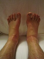
Edema of both legs after walking more than 100 kilometers
-
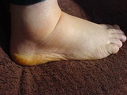
Foot 2 weeks post-surgery
-
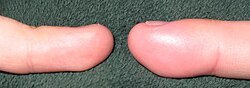
Left and right ring fingers of the same individual. The distal phalanx of the finger on the right exhibits edema due to acute paronychia.


Generalized
A rise in hydrostatic pressure occurs in cardiac failure. A fall in osmotic pressure occurs in nephrotic syndrome and liver failure.[6]
Causes of edema which are generalized to the whole body can cause edema in multiple organs and peripherally. For example, severe heart failure can cause pulmonary edema, pleural effusions, ascites and peripheral edema. Such severe systemic edema is called anasarca. In rare cases, a Parvovirus B19 infection may cause generalized edemas.[7]
Although a low plasma oncotic pressure is widely cited for the edema of nephrotic syndrome, most physicians note that the edema may occur before there is any significant protein in the urine (proteinuria) or fall in plasma protein level. Most forms of nephrotic syndrome are due to biochemical and structural changes in the basement membrane of capillaries in the kidney glomeruli, and these changes occur, if to a lesser degree, in the vessels of most other tissues of the body. Thus the resulting increase in permeability that leads to protein in the urine can explain the edema if all other vessels are more permeable as well.[8]
As well as the previously mentioned conditions, edemas often occur during the late stages of pregnancy in some women. This is more common with those of a history of pulmonary problems or poor circulation also being intensified if arthritis is already present in that particular woman. Women who already have arthritic problems most often have to seek medical help for pain caused from over-reactive swelling. Edemas that occur during pregnancy are usually found in the lower part of the leg, usually from the calf down.
Hydrops fetalis is a condition in a baby characterized by an accumulation of fluid in at least two body compartments.
Cause
Heart
The pumping force of the heart should help to keep a normal pressure within the blood vessels. But if the heart begins to fail (a condition known as congestive heart failure) the pressure changes can cause very severe water retention. In this condition water retention is mostly visible in the legs, feet and ankles, but water also collects in the lungs, where it causes a chronic cough. This condition is usually treated with diuretics; otherwise, the water retention may cause breathing problems and additional stress on the heart.[9]
Kidneys
Another cause of severe water retention is kidney failure, where the kidneys are no longer able to filter fluid out of the blood and turn it into urine. Kidney disease often starts with inflammation, for instance in the case of diseases such as nephrotic syndrome or lupus. Once again, this type of water retention is usually visible in the form of swollen legs and ankles.
Protein
Protein attracts water and plays an important role in water balance. In cases of severe protein deficiency, the blood may not contain enough protein to attract water from the tissue spaces back into the capillaries. This is why starvation often shows an enlarged abdomen. The abdomen is swollen with edema or water retention caused by the lack of protein in their diet.
When the capillary walls are too permeable, protein can leak out of the blood and settle in the tissue spaces. It will then act like a magnet for water, continuously attracting more water from the blood to accumulate in the tissue spaces.[10]
Others
Swollen legs, feet and ankles are common in late pregnancy. The problem is partly caused by the weight of the uterus on the major veins of the pelvis. It usually clears up after delivery of the baby, and is mostly not a cause for concern,[11] though it should always be reported to a doctor.
Lack of exercise is another common cause of water retention in the legs. Exercise helps the leg veins work against gravity to return blood to the heart. If blood travels too slowly and starts to pool in the leg veins, the pressure can force too much fluid out of the leg capillaries into the tissue spaces. The capillaries may break, leaving small blood marks under the skin. The veins themselves can become swollen, painful and distorted – a condition known as varicose veins.[12] Muscle action is needed not only to keep blood flowing through the veins but also to stimulate the lymphatic system to fulfil its "overflow" function. Long-haul flights, lengthy bed-rest, immobility caused by disability and so on, are all potential causes of water retention. Even very small exercises such as rotating ankles and wiggling toes can help to reduce it.[13]
Certain medications are prone to causing water retention. These include estrogens, thereby including drugs for hormone replacement therapy or the combined oral contraceptive pill,[14] as well as non-steroidal anti-inflammatory drugs[15] and beta-blockers.[16]
Premenstrual water retention, causing bloating and breast tenderness, is common.[17][18][19]
Mechanism
Six factors can contribute to the formation of edema:
- increased hydrostatic pressure;
- reduced colloidal or oncotic pressure within blood vessels;
- increased tissue colloidal or oncotic pressure;
- increased blood vessel wall permeability (e.g., inflammation);
- obstruction of fluid clearance in the lymphatic system;
- changes in the water retaining properties of the tissues themselves. Raised hydrostatic pressure often reflects retention of water and sodium by the kidneys.[20]
Generation of interstitial fluid is regulated by the forces of the Starling equation.[21] Hydrostatic pressure within blood vessels tends to cause water to filter out into the tissue. This leads to a difference in protein concentration between blood plasma and tissue. As a result, the colloidal or oncotic pressure of the higher level of protein in the plasma tends to draw water back into the blood vessels from the tissue. Starling's equation states that the rate of leakage of fluid is determined by the difference between the two forces and also by the permeability of the vessel wall to water, which determines the rate of flow for a given force imbalance. Most water leakage occurs in capillaries or post capillary venules, which have a semi-permeable membrane wall that allows water to pass more freely than protein. (The protein is said to be reflected and the efficiency of reflection is given by a reflection constant of up to 1.) If the gaps between the cells of the vessel wall open up then permeability to water is increased first, but as the gaps increase in size permeability to protein also increases with a fall in reflection coefficient.
Changes in the variables in Starling's equation can contribute to the formation of edemas either by an increase in hydrostatic pressure within the blood vessel, a decrease in the oncotic pressure within the blood vessel or an increase in vessel wall permeability. The latter has two effects. It allows water to flow more freely and it reduces the colloidal or oncotic pressure difference by allowing protein to leave the vessel more easily.
Another set of vessels known as the lymphatic system acts like an "overflow" and can return much excess fluid to the bloodstream. But even the lymphatic system can be overwhelmed, and if there is simply too much fluid, or if the lymphatic system is congested, then the fluid will remain in the tissues, causing swellings in legs, ankles, feet, abdomen or any other part of the body.[22]
Diagnosis
| Grade[23] | Definition |
|---|---|
| Absent | Absent |
| + | Mild: Both feet / ankles |
| ++ | Moderate: Both feet, plus lower legs, hands or lower arms |
| +++ | Severe: Generalised bilateral pitting edema, including both feet, legs, arms and face |
Cutaneous edema is referred to as "pitting" when, after pressure is applied to a small area, the indentation persists after the release of the pressure. Peripheral pitting edema, as shown in the illustration, is the more common type, resulting from water retention. It can be caused by systemic diseases, pregnancy in some women, either directly or as a result of heart failure, or local conditions such as varicose veins, thrombophlebitis, insect bites, and dermatitis.
Non-pitting edema is observed when the indentation does not persist. It is associated with such conditions as lymphedema, lipedema, and myxedema.
Edema caused by malnutrition defines kwashiorkor, an acute form of childhood protein-energy malnutrition characterized by edema, irritability, anorexia, ulcerating dermatoses, and an enlarged liver with fatty infiltrates.
Treatment
When possible, treatment involves resolving the underlying cause. Many cases of heart or kidney disease, are treated with diuretics.[9]
Treatment may also involve positioning the affected body parts to improve drainage. For example, swelling in feet or ankles may be reduced by having the person lie down in bed or sit with the feet propped up on cushions. Intermittent pneumatic compression can be used to pressurize tissue in a limb, forcing fluids—both blood and lymph—to flow out of the compressed area.
Compression stockings in those with a history of cellulitis decreases the risk of recurrence.[24]
References
- ↑ 1.0 1.1 1.2 1.3 1.4 1.5 1.6 Causes and signs of edema. Institute for Quality and Efficiency in Health Care (IQWiG). 2016. Archived from the original on 2021-08-29. Retrieved 2019-12-08.
- ↑ 2.0 2.1 2.2 2.3 2.4 2.5 2.6 2.7 2.8 "Edema - Cardiovascular Disorders". Merck Manuals Professional Edition. Archived from the original on 13 October 2019. Retrieved 8 December 2019.
- ↑ 3.0 3.1 3.2 "Edema: Causes, Symptoms, Diagnosis & Treatment". familydoctor.org. Archived from the original on 1 August 2019. Retrieved 23 December 2019.
- ↑ Liddell, Henry. "A Greek-English Lexicon, οἴδ-ημα". www.perseus.tufts.edu. Archived from the original on 7 July 2015. Retrieved 8 December 2019.
- ↑ C.Michael Hogan (2008) "Western poison-oak: Toxicodendron diversilobum" Archived July 21, 2009, at the Wayback Machine, GlobalTwitcher, ed. Nicklas Strömberg
- ↑ Renkin EM (1994). "Cellular aspects of transvascular exchange: a 40-year perspective". Microcirculation. 1 (3): 157–67. doi:10.3109/10739689409148270. PMID 8790586.
- ↑ Wiggli B, Imhof E, Meier CA, Laifer G (2013). "Water, water, everywhere. Acute parvovirus B19 infection". Lancet. 381 (9868): 776. doi:10.1016/S0140-6736(12)61894-7. PMID 23472922.
- ↑ Palmer BF, Alpern RJ (1997). "Pathogenesis of edema formation in the nephrotic syndrome". Kidney Int. Suppl. 59: S21–7. PMID 9185099.
- ↑ 9.0 9.1 Casu, Gavino; Merella, Pierluigi (July 2015). "Diuretic Therapy in Heart Failure – Current Approaches". European Cardiology Review. 10 (1): 42–47. doi:10.15420/ecr.2015.10.01.42. ISSN 1758-3756. PMC 6159465. PMID 30310422.
- ↑ Meisenberg, Gerhard; Simmons, William H. (2006). Principles of Medical Biochemistry (2nd ed.). Philadelphia: Elsevier Health Sciences. p. 258. ISBN 978-0-32302-942-1.
- ↑ Heine, R. Phillips; Swamy, Geeta K. "Lower-Extremity Edema During Late Pregnancy". The Merck Manual. Archived from the original on 24 October 2010. Retrieved 9 August 2017.
- ↑ Timby, Barbara Kuhn; Smith, Nancy E. (2006). Introductory Medical-Surgical Nursing (9th ed.). Philadelphia: Lippincott Williams & Wilkins. p. 488. ISBN 978-0-78178-032-2.
- ↑ Zuther, Joachim E. (2005). Lymphedema Management: The Comprehensive Guide for Practitioners (1st ed.). New York: Thieme Medical Publishers. p. 222. ISBN 978-1-58890-284-9.
- ↑ "Estrogens (Conjugated/Equine)". The Merck Manual. Archived from the original on 2 December 2007. Retrieved 9 August 2017.
- ↑ "Medscape Today". Archived from the original on 2003-08-07. Retrieved 2019-12-08.(subscription required)
- ↑ "Beta-Blockers for High Blood Pressure". WebMD. Archived from the original on 17 November 2012. Retrieved 9 August 2017.
- ↑ Lee-Ellen C. Copstead-Kirkhorn; Jacquelyn L. Banasik (25 June 2014). Pathophysiology. Elsevier Health Sciences. pp. 660–. ISBN 978-0-323-29317-4. Archived from the original on 14 April 2021. Retrieved 8 December 2019.
- ↑ Farage MA, Neill S, MacLean AB (2009). "Physiological changes associated with the menstrual cycle: a review". Obstet Gynecol Surv. 64 (1): 58–72. doi:10.1097/OGX.0b013e3181932a37. PMID 19099613.
- ↑ Charlotte Pooler (1 October 2009). Porth Pathophysiology: Concepts of Altered Health States. Lippincott Williams & Wilkins. pp. 1075, 1107. ISBN 978-1-60547-781-7. Archived from the original on 16 December 2019. Retrieved 8 December 2019.
- ↑ Kumar; Abbas; Fausto (1999). Pathologic Basis of Disease (7th ed.). Elsevier Saunders. p. 122. ISBN 0-7216-0187-1.
- ↑ Boron W.F., Boulpaep E.L. (2012.) Medical Physiology: A Cellular and Molecular Approach, 2e. Saunders/Elsevier, Philadelphia, PA.
- ↑ Rubin, Emanuel (2008). Essentials of Rubin's Pathology (5th ed.). Philadelphia: Lippincott Williams & Wilkins. p. 124. ISBN 978-0-78177-324-9.
- ↑ Nutrition in Emergencies > Measuring œdema Archived 2017-02-18 at the Wayback Machine. Erin Boyd, reviewed by Diane Holland, Nutrition in Emergencies Unit, UNICEF. Retrieved Nov 2012
- ↑ Ton, Joey (1 November 2021). "#301 Under Pressure: Compression stockings for recurrent cellulitis?". CFPCLearn. Archived from the original on 2 February 2023. Retrieved 14 June 2023.
External links
| Classification | |
|---|---|
| External resources |