Cryoglobulinemic vasculitis

From WikiProjectMed
Jump to navigation Jump to search
Cryoglobulinemic vasculitis
Other names: Essential mixed cryoglobulinemia, Primary cryoglobulinemia[1]
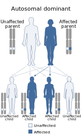
Cryoglobulinemic vasculitis is inherited in an autosomal dominant manner
SpecialtyDermatology

Cryoglobulinemic vasculitis is a form of inflammation affecting the blood vessels caused by the deposition of abnormal proteins called cryoglobulins in the blood vessels, in patients affected by mixed cryoglobulinemia (type 2 and 3), since simple or type 1 cryoglobulinemia does not cause complement activation (but a hyperviscosity syndrome only). Cryoglobulinemic vasculitis affects the skin and causes a rash in roughly 15% of people with detectable circulating cryoglobulin proteins.[2]: 835  Additionally, the kidneys may be affected by this form of vasculitis resulting in membranoproliferative glomerulonephritis. Fevers, painful muscles and joints, and peripheral nerve damage are other common manifestations of cryoglobulinemic vasculitis. Due to deposition of complement (in particular, C4), low levels of circulating complement factors may be seen.
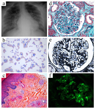
Widespread fulminant cryoglobulinemic vasculitis is defined as involvement of the skin and at least two other organs, such as illustrated by the following case. A 50-year-old woman presented with fever, bilateral purpuric lesions, dyspnea, and acute kidney injury. The bronchoalveolar lavage showed severe hemorrhage with predominance of macrophages and neutrophils (a, May-Grünwald stain, x200). The skin biopsy revealed typical features of leukocytoclastic vasculitis with intraluminal thrombosis and fibrinoid-type necrosis associated with infiltration by neutrophils of the vessel walls in the dermis (b, hematoxylin-eosin-saffron stain, x200). The kidney biopsy displayed membranoproliferative glomerulonephritis. The glomeruli showed mesangial expansion by increased mesangial cell number and matrix, and some intracapillary "protein thrombi" (c, Masson’s trichrome stain, x400). A mild endocapillary hypercellularity and duplication of the glomerular basement membrane with “double contours” were also observed (d, Jones stain, x400). Immunofluorescence study showed granular IgG-positive deposits with lambda light-chain isotype restriction localized to the glomerular capillary wall, mesangium, and intracapillary thrombi (e, IgG Immunofluorescence staining, x400, lambda staining not shown). IgM, IgA, and kappa staining were all negative (data not shown). Lambda staining was comparable to IgG staining (data not shown), which was consistent with renal involvement of type I cryoglobulinemia (IgG lambda). Finally, the diagnosis was widespread cryoglobulinemic vasculitis with skin, lung, and kidney involvement secondary to monoclonal gammopathy of undetermined significance associated with Sjögren’s syndrome.

See also

References

  1. RESERVED, INSERM US14-- ALL RIGHTS. "Orphanet: Cryoglobulinemic vasculitis". www.orpha.net. Archived from the original on 20 January 2021. Retrieved 19 April 2019.
  2. James, William D.; Berger, Timothy G.; et al. (2006). Andrews' Diseases of the Skin: clinical Dermatology. Saunders Elsevier. ISBN 978-0-7216-2921-6.

External links

Classification
External resources