Cervical cancer staging is the assessment of cervical cancer to determine the extent of the disease. This is important for determining disease prognosis and treatment. Cancer staging generally runs from stage 0, which is pre-cancerous or non-invasive, to stage IV, in which the cancer has spread throughout a significant part of the body.[1]
FIGO guidelines suggest that lower staging should be assigned whenever possible. Staging is determined after all imaging and pathology reports have been collected and interpreted. The stage of cervical cancer cannot be altered at recurrence.[4]
Imaging
The use of imaging can provide more information regarding prognosis, which informs treatment. The goal of staging is to identify the most appropriate treatment method and to minimize dual therapy, which can significantly impact morbidity. The imaging modality used for diagnosis should always be noted. Imaging modalities include ultrasound, MRI, CT, and positron emission tomography (PET).[5]
For primary tumors over 10 mm, MRI is the best method of radiologic assessment.[6] For nodal metastasis over 10 mm, PET-CT is the most accurate modality for diagnosis, as this can differentiate large lymph nodes that are not metastatic (for example, those that are enlarged due to inflammation or infection) from lymph nodes that are enlarged due to metastases. The diagnosis of nodal metastasis can be supplemented with surgical assessment via minimally invasive surgery or laparotomy to collect a biopsy or perform a fine needle aspiration.[7]
Pathology
As with imaging, pathologic methods used for diagnosis should always be noted for future evaluation. All cancers must be confirmed by microscopic examination.[5]
Cancers can be histopathologically graded as follows:
GX: Grade cannot be assessed
G1: Well differentiated
G2: Moderately differentiated
G3: Poorly or undifferentiated
Sample collection
For microinvasive disease, clinicians should collect a specimen using a loop electrosurgical excision procedure (LEEP) or a cone biopsy. If a patient has a visible lesion, a punch biopsy may be attempted. If the specimen is not satisfactory, a small loop biopsy or cone may be required.[citation needed]
Stages
Stage 0
Diagram showing extent of cervical cancer tumor in blue corresponding to sub-stages IA1 and IA2.[9]The carcinoma is confined to the surface layer (cells lining) of the cervix. Also called carcinoma in situ (CIS).[4]
Stage I
The carcinoma has grown into the cervix, but has not spread beyond it (extension to the corpus would be disregarded). Stage One is subdivided as follows:[4]
IA: Invasive carcinoma which can be diagnosed only by microscopy on a LEEP or cone biopsy specimen, or on a trachelectomy or hysterectomy specimen, with deepest invasion <5 mm from the base of the epithelium. It can originate from squamous or glandular epithelium. The margins of the specimen should be reported to be negative for disease. Lymphovascular involvement must be noted to inform the treatment plan, although it does not change stage designation.[5]
IA1: Measured stromal invasion <3.0 mm
IA2: Measured stromal invasion ≥3.0 mm and <5 mm
IB: Invasive carcinoma with measured deepest invasion ≥5 mm, limited to the cervix. The lesions are clinically visible, which differentiates IB from IA. If the margins of the cone biopsy are positive for invasive disease, Stage IB1 is automatically assigned. As with Stage IA, the involvement of vascular or lymphatic spaces does not change the staging, but should be noted to inform the treatment plan.
IB1: Invasive carcinoma ≥5 mm depth of invasion and <2 cm in greatest dimension
IB2: Invasive carcinoma ≥2 cm and <4 cm in greatest dimension
IB3: Invasive carcinoma ≥4.0 cm in greatest dimensionDiagram of sub-stage IB1.[9]Diagram of sub-stage IB2.[9]Diagram of sub-stage IB3.[9]
Stage II
Cervical carcinoma invades beyond the uterus, but not to the pelvic wall or to the lower third of the vagina[4]
IIA2: Tumor ≥4.0 cm in greatest dimensionDiagram of sub-stage IIA.[10]
IIB: With parametrial invasionDiagram of sub-stage IIB.[10]
Stage III
The carcinoma involves the lower third of the vagina and/or extends to the pelvic wall and/or causes hydronephrosis or non‐functioning kidney and/or involves pelvic and/or para-aortic lymph nodes.[5]
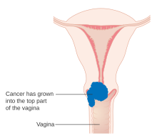
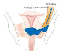
IIIA: Carcinoma involves the lower third of the vagina, with no extension to the pelvic wall.Diagram of sub-stage IIIA.[11]IIIB: Extension to the pelvic wall and/or hydronephrosis or non‐functioning kidney.Diagram of sub-stage IIIB.[11]
IIIC: Involvement of pelvic and/or para-aortic lymph nodes, irrespective of tumor size and extent
IIIC1: Pelvic lymph node metastasis onlyDiagram of sub-stage IIIC1.[11]
IIIC2: Para‐aortic lymph node metastasisDiagram of sub-stage IIIC2.[11]
Stage IV
The carcinoma has extended beyond the true pelvis or has involved (biopsy proven) the mucosa of the bladder or rectum. A bullous edema, as such, does not permit a case to be allotted to Stage IV[4]
IVA: Spread of the growth to adjacent organsDiagram of sub-stage IVA.[12]
IVB: Spread to distant organsDiagram of sub-stage IVB.[12]
History of cervical cancer staging
The drive to develop a staging for gynecological malignancies, including cancer of the cervix, was the need to have a uniform method to describe the extent of the disease. Comparing outcomes from different treatments could only be possible if the comparison were made for groups of patients with a similar degree of disease burden.[13]
The most recent updates to cervical cancer staging included in the 2018 edition marked a departure from a system based mainly on clinical evaluation to one that allows imaging and pathological methods to help determine tumor size and extent of disease to assign the stage. A major topic of debate was the impact that newer diagnostic modalities would have on low- and middle-income countries, which bear most of the disease burden.[15] The final recommendations make findings from imaging and pathology optional for staging rather than a requirement.[citation needed]
References
↑"Staging". National Cancer Institute. 9 March 2015. Archived from the original on 2015-03-19. Retrieved 2018-11-07.