Calciphylaxis
| Calciphylaxis | |
|---|---|
| Other names: Calcific uremic arteriolopathy (CUA)[1] | |
 | |
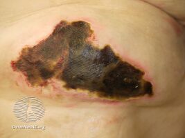
| Calciphylaxis on the abdomen of a person with end stage kidney disease. Markings are in cm. | |
| Symptoms | Severe pain and skin changes[1] |
| Complications | Depression, sepsis[1] |
| Causes | Unknown[2] |
| Risk factors | End-stage kidney disease, earlier stage chronic kidney disease, acute kidney injury[1] |
| Diagnostic method | Based on symptoms, skin biopsy[2] |
| Differential diagnosis | Antiphospholipid syndrome, cholesterol embolism, vasculitis, warfarin necrosis[2] |
| Treatment | Increased dialysis frequency, stopping calcium supplements, sodium thiosulfate[2] |
| Prognosis | 1 year survival < 50%[1] |
| Frequency | 4 to 400 per 10,000 on dialysis/year[2] |
Calciphylaxis is calcification of small blood vessels located within fatty tissue and the deeper layer of the skin.[1] Symptoms generally include severe pain and skin changes.[1] Often their are multiple areas of skin involvement, beginning as areas of thickening, that become purple in color, followed by the formation of an ulcer.[1] Complications may include depression and sepsis.[1]
It most commonly occurs in those with end-stage kidney disease; though can occur in earlier stage chronic kidney disease or acute kidney injury.[1] Rarely it may occur in those with normal kidney function.[1] Other risk factors include obesity, diabetes, cancer, certain medications including warfarin and vitamin D, and areas of tissue injury.[2] The underlying mechanism involves narrowing of small blood vessels followed by blood clots and the death of skin cells due to too little blood flow.[2] Diagnosis may be made based on symptoms; though can be confirmed by skin biopsy.[2]
Treatment may involved increased dialysis frequency, stopping calcium supplements, and sodium thiosulfate.[2] For those with high parathyroid hormone, cinacalcet should be used instead of activated vitamin D.[2] Other important efforts include wound care and pain management.[2] Hyperbaric oxygen therapy may help some people.[2] Outcomes are generally poor, with a typical life expectancy of less than one year.[1] The cause of death is most often sepsis.[2]
Calciphylaxis is rare, affecting about 4 to 400 per 10,000 people on dialysis per year.[2] Women are more commonly affected than men and all ages may be involved.[3] The condition was first described in 1898 by White and Bryant.[3] The importance of the condition was determined in 1976 by Gipstein.[3]
Signs and symptoms
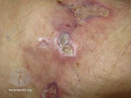
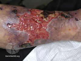
The first skin changes in calciphylaxis lesions are mottling of the skin and induration in a livedo reticularis pattern. As tissue thrombosis and infarction occurs, a black, leathery eschar in an ulcer with adherent black slough are found. Surrounding the ulcers is usually a plate-like area of indurated skin.[4] These lesions are always extremely painful and most often occur on the lower extremities, abdomen, buttocks, and penis. Because the tissue has infarcted, wound healing seldom occurs, and ulcers are more likely to become secondarily infected. Many cases of calciphylaxis end with systemic bacterial infection and death.[5]
Calciphylaxis is characterized by the following histologic findings:
- systemic medial calcification of the arteries, i.e. calcification of tunica media. Unlike other forms of vascular calcifications (e.g., intimal, medial, valvular), calciphylaxis is characterized also by
- small vessel mural calcification with or without endovascular fibrosis, extravascular calcification and vascular thrombosis, leading to tissue ischemia (including skin ischemia and, hence, skin necrosis).
-
Calciphylaxis
-
Calciphylaxis
-
Calciphylaxis
.jpg)
.jpg)
.jpg)
Heart of stone
Severe forms of calciphylaxis may cause diastolic heart failure from cardiac calcification, called heart of stone.[6]
Cause
The cause of calciphylaxis is unknown. It does not seem to be an immune type reaction. In other words, calciphylaxis is not a hypersensitivity reaction (i.e., allergic reaction) leading to sudden local calcification. Clearly, additional factors are involved in calciphylaxis. It is also known as calcific uremic arteriolopathy; however, the disease is not limited to patients with kidney failure. The current belief is that in end-stage kidney disease, abnormal calcium and phosphate homeostasis result in the deposition of calcium in the vessels, also known as metastatic calcification. Once the calcium has been deposited, a thrombotic event occurs within the lumen of these vessels, resulting in tissue infarction. It is unknown what the triggers are that cause the thrombotic and ischemic event.[7] Reported risk factors include female sex, obesity, elevated calcium-phosphate product, medications such as warfarin, vitamin D derivatives e.g. calcitriol, calcium-based binders, or systemic steroids, protein C or S deficiency, low blood albumin levels, and diabetes mellitus.[8]
Diagnosis
There is no diagnostic test for calciphylaxis. The diagnosis is a clinical one. The characteristic lesions are the ischemic skin lesions (usually with areas of skin necrosis). The necrotic skin lesions (i.e. the dying or already dead skin areas) typically appear as violaceous (dark bluish purple) lesions and/or completely black leathery lesions. They can be extensive. The suspected diagnosis can be supported by a skin biopsy. It shows arterial calcification and occlusion in the absence of vasculitis. Sometimes the bone scintigraphy can show increased tracer accumulation in the soft tissues.[9] In certain patients, anti-nuclear antibody may play a role.[10]
Treatment
The optimal treatment is prevention. Rigorous and continuous control of phosphate and calcium balance most probably will avoid the metabolic changes which may lead to calciphylaxis.
There is no specific treatment. Of the treatments that exist, none are standard of care. Acceptable treatment could include:
- Dialysis (the number of sessions may be increased)
- Intensive wound care
- Clot-dissolving agents (tissue plasminogen activator)
- Hyperbaric oxygen[11]
- Maggot larval debridement
- Adequate pain control
- Correction of the underlying plasma calcium and phosphorus abnormalities (lowering the Ca x P product below 55 mg2/dL2)
- Sodium thiosulfate
- Avoiding (further) local tissue trauma (including avoiding all subcutaneous injections, and all not-absolutely-necessary infusions and transfusions)
- Urgent parathyroidectomy: The efficacy of this measure remains uncertain although calciphylaxis is associated with frank hyperparathyroidism. Urgent parathyroidectomy may benefit those patients who have uncontrollable plasma calcium and phosphorus concentrations despite dialysis. Also, cinacalcet can be used and may serve as an alternative to parathyroidectomy.
- Patients who receive kidney transplants also receive immunosuppression. Considering lowering the dose of or discontinuing the use of immunosuppressive drugs in people who have received kidney transplants and continue to have persistent or progressive calciphylactic skin lesions can contribute to an acceptable treatment of calciphylaxis.
- A group has reported plasma exchange effective and propose a serum marker and perhaps mediator (calciprotein particles)[12]
Prognosis
Response to treatment is not guaranteed. Also, the necrotic skin areas may get infected, and this then may lead to sepsis in some patients. Overall, the clinical prognosis remains poor.
Epidemiology
Calciphylaxis most commonly occurs in patients with end-stage renal disease who are on hemodialysis or who have recently received a renal (kidney) transplant. Yet calciphylaxis does not occur only in end-stage renal disease patients. When reported in patients without end-stage renal disease, it is called non-uremic calciphylaxis by Nigwekar et al.[13] Non-uremic calciphylaxis has been observed in patients with primary hyperparathyroidism, breast cancer (treated with chemotherapy), liver cirrhosis (due to hazardous alcohol use), cholangiocarcinoma, Crohn's disease, rheumatoid arthritis (RA), and systemic lupus erythematosus (SLE).
References
- ↑ 1.00 1.01 1.02 1.03 1.04 1.05 1.06 1.07 1.08 1.09 1.10 1.11 Nigwekar, SU; Thadhani, R; Brandenburg, VM (May 2018). "Calciphylaxis". New England Journal of Medicine. 378 (18): 1704–1714. doi:10.1056/NEJMra1505292. PMID 29719190.
- ↑ 2.00 2.01 2.02 2.03 2.04 2.05 2.06 2.07 2.08 2.09 2.10 2.11 2.12 2.13 Westphal, SG; Plumb, T (January 2022). "Calciphylaxis". PMID 30085562.
{{cite journal}}: Cite journal requires|journal=(help) - ↑ 3.0 3.1 3.2 Herndon, David N. (15 June 2012). Total Burn Care E-Book: Expert Consult - Online. Elsevier Health Sciences. p. 480. ISBN 978-1-4557-3797-0. Archived from the original on 14 October 2022. Retrieved 13 October 2022.
- ↑ Zhou Qian; Neubauer Jakob; Kern Johannes S; Grotz Wolfgang; Walz Gerd; Huber Tobias B (2014). "Calciphylaxis". The Lancet. 383 (9922): 1067. doi:10.1016/S0140-6736(14)60235-X. PMID 24582472.
- ↑ Wolff, Klaus; Johnson, Richard; Saavedra, Arturo (2013-03-06). Fitzpatrick's Color Atlas and Synopsis of Clinical Dermatology (7th ed.). McGraw Hill. p. 429. ISBN 978-0-07-179302-5.
- ↑ Heart of Stone Archived 2007-03-10 at the Wayback Machine - CINDY W. T OM, MD, ANDDEEPAKR. TALREJA, MD. Division of Cardiovascular Diseases, Mayo Clinic College of Medicine, Rochester, Minn
- ↑ Wilmer, William; Magro, Cynthia (2002). "Calciphylaxis: Emerging Concepts in Prevention, Diagnosis, and Treatment". Seminars in Dialysis. 15 (3): 172–186. doi:10.1046/j.1525-139X.2002.00052.x. PMID 12100455.
- ↑ Arseculeratne, G; Evans, AT; Morley, SM (2006). "Calciphylaxis – a topical overview". Journal of the European Academy of Dermatology and Venereology. 20 (5): 493–502. doi:10.1111/j.1468-3083.2006.01506.x. PMID 16684274.
- ↑ Araya CE, Fennell RS, Neiberger RE, Dharnidharka VR (2006). "Sodium thiosulfate treatment for calcific uremic arteriolopathy in children and young adults". Clin J Am Soc Nephrol. 1 (6): 1161–6. doi:10.2215/CJN.01520506. PMID 17699342. Archived from the original on 2009-11-19. Retrieved 2021-03-30.
- ↑ Rashid RM, Hauck M, Lasley M (Nov 2008). "Anti-nuclear antibody: a potential predictor of calciphylaxis in non-dialysis patients". J Eur Acad Dermatol Venereol. 22 (10): 1247–8. doi:10.1111/j.1468-3083.2008.02606.x. PMID 18422539.
- ↑ Edsell ME, Bailey M, Joe K, Millar I (2008). "Hyperbaric oxygen therapy in the treatment of skin ulcers due to calcific uraemic arteriolopathy: experience from an Australian hyperbaric unit". Diving and Hyperbaric Medicine. 38 (3): 139–44. Archived from the original on 2014-10-10. Retrieved 2013-04-02.
- ↑ Cai MM, Smith ER, Brumby C, McMahon LP, Holt SG (2013). "Fetuin-A-containing calciprotein particle levels can be reduced by dialysis, sodium thiosulphate and plasma exchange. Potential therapeutic implications for calciphylaxis?". Nephrology (Carlton). 18: 724–7. doi:10.1111/nep.12137. PMID 24571743.
{{cite journal}}: CS1 maint: multiple names: authors list (link) - ↑ Nigwekar SU, Wolf M, Sterns RH, Hix JK (Jul 2008). "Calciphylaxis from nonuremic causes: a systematic review". Clin J Am Soc Nephrol. 3 (4): 1139–43. doi:10.2215/cjn.00530108. PMC 2440281. PMID 18417747.
Further reading
- Weenig RH (2008). "Pathogenesis of calciphylaxis: Hans Selye to nuclear factor kappa-B". J. Am. Acad. Dermatol. 58 (3): 458–71. doi:10.1016/j.jaad.2007.12.006. PMID 18206262.
- Weenig RH, Sewell LD, Davis MD, McCarthy JT, Pittelkow MR (2007). "Calciphylaxis: natural history, risk factor analysis, and outcome". J. Am. Acad. Dermatol. 56 (4): 569–79. doi:10.1016/j.jaad.2006.08.065. PMID 17141359.
- Li JZ, Huen W (2007). "Images in clinical medicine. Calciphylaxis with arterial calcification". N. Engl. J. Med. 357 (13): 1326. doi:10.1056/NEJMicm060859. PMID 17898102.
External links
| Classification | |
|---|---|
| External resources |