Bone tumor
| Bone tumor | |
|---|---|
  | |
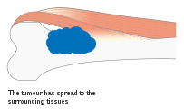
| Top : non-cancerous bone tumor, bottom: cancerous bone tumor | |
| Specialty | Orthopedics |
| Symptoms | Lump, pain, neurological signs, unexplained broken bone,[1] fatigue, fever, weight loss, anemia and nausea.[2][3] Sometimes no symptoms.[1] |
| Types | Noncancerous (benign) or cancerous (malignant)[4] |
| Diagnostic method | Medical imaging, biopsy[4] |
| Prognosis | Varies with type[1] |
| Frequency | Common[1] |
A bone tumor is an abnormal growth of tissue in bone, traditionally classified as noncancerous (benign) or cancerous (malignant).[1][4] The classification of bone tumors was revised by the World Health Organization (WHO) in 2020,[5] and categories include cartilage tumors, tumors of connective tissue, blood vessels, and bone forming tumors.[6]
A bone tumor may present with a lump, pain, or neurological features from compression.[4] There may be an unexplained broken bone; with little or no trauma.[1][2] Other symptoms may include fatigue, fever, weight loss, anemia and nausea.[2][3] Sometimes there are no symptoms and the tumour is found when investigating another problem.[2][3]
For most bone tumors, the cause is unknown.[1] Cancerous bone tumors rarely begin in the bone itself, and usually originate from a cancer in another part of the body such as cancer of the lung, breast, thyroid, kidney or prostate.[1][2] Some people such as those with Paget's disease or longterm bone infection may have an increased risk of developing a cancerous bone tumor.[1]
Diagnosis is generally by X-ray and other radiological tests such as CT scan, MRI, PET scan and bone scintigraphy.[4] Blood tests might include a complete blood count, inflammatory markers, serum electrophoresis, PSA, kidney function and liver function.[4] Urine may be sent for Bence Jones protein.[4] For confirmation of diagnosis, a biopsy for histological evaluation might be required.[4][2]
Treatment of bone tumors is dependent on the type of tumor.[2] Generally, noncancerous bone tumors may be observed for changes and surgery offered if there is pain or pressure effects on neighbouring body parts.[4] A surgical resection with or without cytotoxic drugs may be considered.[4]
Bone tumors arising from bone are rare and account for around 0.2% of all human tumors.[6] The most common bone tumor is a benign non-ossifying fibroma.[1] Average five-year survival in the United States after being diagnosed with bone and joint cancer is 67%.[7] The earliest known bone tumor is dated 1.6 to 1.8 million years ago.[8]
Classification

Bone tumors are traditionally classified as noncancerous (benign) or cancerous (malignant).[4] Several features of bone tumors and soft tissue tumors overlap.[6] Their classification was revised by the World Health Organization (WHO) in 2020.[5] This newer classification divides bone tumors into tumors of cartilage, bone forming tumors, fibrogenic tumors, vascular tumors of bone, osteoclastic giant cell-rich tumors, notochordal tumors, other mesenchymal tumors of bone, and vascular tumors of bone.[1]
Noncancerous
Noncancerous (benign) bone tumors can be abnormal growths of bone, cartilage, fibrous tissue and blood vessels.[9] The most common bone tumor is a non-ossifying fibroma.[1][10] Others include chondroblastoma, osteoid osteoma, enchondroma, giant cell tumor of bone,[4] unicameral bone cyst, eosinophilic granuloma haemangioma, osteoblastoma and aneurysmal bone cyst.[9]
Cancerous
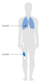
Cancerous bone tumors are commonly secondary; that is they originate from a cancer in another part of the body.[2][9] They are more likely in long bones, spine and in older people, and are generally due to spread from cancers of other organs.[11]
Primary malignant bone tumors include osteosarcoma, chondrosarcoma, Ewing's sarcoma, spindle cell sarcoma, and multiple myeloma.[2]
Signs and symptoms
Clinical features of a bone tumor depend on the type of tumor and which part of which bone is affected.[2][12] Symptoms and signs usually result from the pressure effect of the tumor.[4]
There may be a lump, with or without pain.[4] Pain may increase with the growth of the tumor and may be worse at night and at rest.[4][3] A bone tumor might present with an unexplained broken bone; with little or no trauma.[2] Additional symptoms may include fatigue, fever, weight loss, anemia and nausea.[2][3] If the tumor presses a nerve, neurological features may be present.[4] Sometimes there are no symptoms and the tumour is found when investigating another problem.[2][3]
Causes
For most bone tumors, the cause is unknown.[1] Most cancerous tumors that originate in bone, occur spontaneously.[1] Some occur within a noncancerous condition of bone such as Paget's disease or longterm bone infection.[1] Some non-cancerous bone tumors increase the risk of having cancerous bone tumor.[1]
Cancers that spread to bone include cancers of the lung, breast, thyroid, kidney and prostate.[2] In younger people, spread to bone is more likely from cancers involving the nervous system, kidneys and soft tissue.[11] Rarely, primary bone cancers such as osteosarcoma may also spread to other bones.{{citation needed|date=April 2021
Diagnosis
A bone tumor may be felt on examination, following which a plain X-ray is usually carried out.[4][13] Imaging is interpreted with the location of the lesion and the person's age being taken into account.[14]
Blood tests might include a complete blood count, inflammatory markers, serum electrophoresis, PSA, kidney function and liver function.[4] Urine may be sent for Bence Jones protein.[4] Other tests that might be requested include a CT scan, MRI, PET scan and bone scintigraphy. For confirmation of diagnosis, a biopsy for histological evaluation might be required, using either a needle or by incision (open biopsy).[4][2]
-

X-ray appearances of different types of bone tumors in < 30 years.
-

X-ray appearances of different types of bone tumors in > 30 years.
-
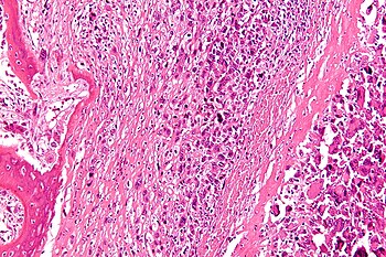
Micrograph of an osteosarcoma, a malignant primary bone tumor.


Staging
-

Stage 1A bone cancer
-

Stage 1B bone cancer
-

Stage 2A bone cancer
-
Stage 2B bone cancer
-
Stage 3 bone cancer


Treatment
Treatment of bone tumors is dependent on the type of tumor.[2] Where available, people with bone tumors are treated at a specialist centre which have surgeons, radiologists, pathologists, oncologists and other support staff.[4] Generally, noncancerous bone tumors may be observed for changes and surgery offered if there is pain or pressure effects on neighbouring body parts. A surgical resection with or without cytotoxic drugs may be considered.[4]
Chemotherapy and radiotherapy
Different regimes of multi-agent adjunct chemotherapy exist and has shown improved survivals with osteosarcoma. Several cycles of neo-adjuvunct chemotherapy may be required to shrink a tumor, such as a Ewing's tumor, prior to surgery.[4] In cases of cancer spread to bone from elsewhere, a broken bone or where surgery is limited due to the margin of the tumor, radiotherapy may help in some types of bone tumor.[4] Although radiotherapy may reduce the extent of surgery, it may cause a greater chance of wound complications.[4]
Medication
One of the major concerns is bone density and bone loss. Non-hormonal bisphosphonates increase bone strength and are available as once-a-week prescription pills. Metastron also known as strontium-89 chloride is an intravenous medication given to help with the pain and can be given in three-month intervals. Generic Strontium Chloride Sr-89 Injection UPS, manufactured by Bio-Nucleonics Inc., it is the generic version of Metastron.[15]
Surgery
Treatment for some bone cancers may involve surgery, such as limb amputation, or limb sparing surgery (often in combination with chemotherapy and radiation therapy). Limb sparing surgery, or limb salvage surgery, means the limb is spared from amputation. Instead of amputation, the affected bone is removed and replaced in one of two ways: (a) bone graft, in which bone is taken from elsewhere on the body or (b) artificial bone is put in. In upper leg surgeries, limb salvage prostheses are available.
The other surgery is called Van Nes rotation or rotationplasty which is a form of amputation, in which the person's foot is turned upwards in a 180-degree turn and the upturned foot is used as a knee.
The most radical of amputations is hemicorporectomy (translumbar or waist amputation) which removes the legs, the pelvis, urinary system, excretory system and the genital area (penis/testes in males and vagina/vulva in females). This operation is done in two stages. First stage is doing the colostomy and the urinary conduit, the second stage is the amputation. This is a mutilating operation and is only done as a last resort (e.g. when even pelvic exenteration does not work or in cases of advanced pelvic/reproductive cancers)
Thermal ablation
CT guided radiofrequency ablation is a less invasive alternative to surgical removal in the care of benign bone tumors, most notably osteoid osteomas. In this technique, which can be performed under precedural sedation, a RF probe is introduced into the tumor nidus through a cannulated needle under CT guidance and heat is applied locally to destroy tumor cells. Since the procedure was first introduced for the treatment of osteoid osteomas in the early 1990s,[16] it has been shown in numerous studies to be less invasive and expensive, to result in less bone destruction and to have equivalent safety and efficacy to surgical techniques, with 66 to 96% of patients reporting freedom from symptoms.[17][18][19] While initial success rates with RFA are high, symptom recurrence after RFA treatment has been reported, with some studies demonstrating a recurrence rate similar to that of surgical treatment.[20]
Thermal ablation techniques are also increasingly being used in the palliative treatment of painful metastatic bone disease. Currently, external beam radiation therapy is the standard of care for patients with localized bone pain due to metastatic disease. Although the majority of patients experience complete or partial relief of pain following radiation therapy, the effect is not immediate and has been shown in some studies to be transient in more than half of patients.[21] For patients who are not eligible or do not respond to traditional therapies ( i.e. radiation therapy, chemotherapy, palliative surgery, bisphosphonates or analgesic medications), thermal ablation techniques have been explored as alternatives for pain reduction. Several multi-center clinical trials studying the efficacy of RFA in the treatment of moderate to severe pain in patients with metastatic bone disease have shown significant decreases in patient reported pain after treatment.[22][23] These studies are limited however to patients with one or two metastatic sites; pain from multiple tumors can be difficult to localize for directed therapy. More recently, cryoablation has also been explored as a potentially effective alternative as the area of destruction created by this technique can be monitored more effectively by CT than RFA, a potential advantage when treating tumors adjacent to critical structures.[24]
Outcomes
The outlook depends on the type of tumor. The outcome is expected to be good for people with noncancerous (benign) tumors, although some types of benign tumors may eventually become cancerous (malignant). With malignant bone tumors that have not spread, most patients achieve a cure, but the cure rate depends on the type of cancer, location, size, and other factors.[citation needed]
Epidemiology
The actual incidence of bone tumors is unknown, as many have no symptoms, and remain undiagnosed.[1] The increased reporting of bone tumors coincides with improved medical imaging.[1] Bone tumors that originate from bone are generally common, but serious types are rare.[1] Non-ossifying fibroma is the most common bone tumor.[1] Tumors arising from cartilage are relatively common findings on knee MRI and have a prevalenee of around 2.8%.[1] Osteosarcoma is the most common cancerous bone tumor, after malignant myeloma.[1]
Bone tumors that originate from bone account for around 0.2% of all tumors.[6] Average five-year survival in the United States after being diagnosed with bone and joint cancer is 67%.[7]
History
The earliest known bone tumor was an osteosarcoma in a foot bone belonging to a person who died in Swartkrans Cave, South Africa, between 1.6 and 1.8 million years ago.[8]
Other animals
Bones are a common site for tumors in cats and dogs.[25]
References
- ↑ 1.00 1.01 1.02 1.03 1.04 1.05 1.06 1.07 1.08 1.09 1.10 1.11 1.12 1.13 1.14 1.15 1.16 1.17 1.18 1.19 1.20 1.21 Soft Tissue and Bone Tumours: WHO Classification of Tumours. International Agency for Research on Cancer. 2020. pp. 338–344. ISBN 978-92-832-4502-5. Archived from the original on 2021-06-13. Retrieved 2021-05-08.
- ↑ 2.00 2.01 2.02 2.03 2.04 2.05 2.06 2.07 2.08 2.09 2.10 2.11 2.12 2.13 2.14 2.15 "Bone Tumor - Types and Treatments - OrthoInfo - AAOS". www.orthoinfo.org. Archived from the original on 20 April 2021. Retrieved 20 April 2021.
- ↑ 3.0 3.1 3.2 3.3 3.4 3.5 "Questions and Answers about Bone Cancer" (PDF). Centers for Disease Control and Prevention. Archived (PDF) from the original on 2 October 2018. Retrieved 18 April 2012.
- ↑ 4.00 4.01 4.02 4.03 4.04 4.05 4.06 4.07 4.08 4.09 4.10 4.11 4.12 4.13 4.14 4.15 4.16 4.17 4.18 4.19 4.20 4.21 4.22 4.23 4.24 Maruthainar, Nimalan; Bhumbra, Rej; Cannon, Steve (2018). "7. Orthopaedic oncology". In Ramachandran, Manoj (ed.). Basic Orthopaedic Sciences (2nd ed.). CRC Press. pp. 105–121. ISBN 978-1-4441-2098-1. Archived from the original on 2021-06-13. Retrieved 2021-04-25.
- ↑ 5.0 5.1 Anderson, William J.; Doyle, Leona A. (2021). "Updates from the 2020 World Health Organization Classification of Soft Tissue and Bone Tumours". Histopathology. 78 (5): 644–657. doi:10.1111/his.14265. ISSN 1365-2559. Archived from the original on 2021-06-27. Retrieved 2021-05-08.
- ↑ 6.0 6.1 6.2 6.3 Choi, Joon Hyuk; Ro, Jae Y. (1 May 2021). "The 2020 WHO Classification of Tumors of Bone: An Updated Review". Advances in Anatomic Pathology. 28 (3): 119–138. doi:10.1097/PAP.0000000000000293. ISSN 1533-4031. PMID 33480599. Archived from the original on 23 May 2021. Retrieved 16 May 2021.
- ↑ 7.0 7.1 "SEER Stat Fact Sheets: Bone and Joint Cancer". NCI. Archived from the original on 20 April 2021. Retrieved 20 April 2021.
- ↑ 8.0 8.1 Strauss, Mark (28 July 2016). "Earliest Human Cancer Found in 1.7-Million-Year-Old Bone". Culture. Archived from the original on 27 June 2021. Retrieved 27 June 2021.
- ↑ 9.0 9.1 9.2 "Bone tumours. What are Bone Tumours?. Patient". patient.info. Archived from the original on 24 April 2021. Retrieved 24 April 2021.
- ↑ Ahn, Leah; O'Donnell, Patrick. "Non-Ossifying Fibroma - Pathology - Orthobullets". www.orthobullets.com. Archived from the original on 5 May 2021. Retrieved 5 May 2021.
- ↑ 11.0 11.1 WHO Classification of Tumours Editorial Board, ed. (2020). "Bone metastases". Soft Tissue and Bone Tumours: WHO Classification of Tumours. Vol. 3 (5th ed.). Lyon (France): International Agency for Research on Cancer. pp. 483–485. ISBN 978-92-832-4503-2. Archived from the original on 2021-06-13. Retrieved 2021-05-08.
- ↑ Murali, Sundaram; Ilaslan, Hakan; Holden, Darlene M. (2015). "2. An imaging approach to bone tumors". In Santini-Araujo, Eduardo; Kalil, Ricardo K.; Bertoni, Franco; Park, Yong-Koo (eds.). Tumors and Tumor-Like Lesions of Bone: For Surgical Pathologists, Orthopedic Surgeons and Radiologists. Springer. pp. 15–56. ISBN 978-1-4471-6577-4. Archived from the original on 2021-06-27. Retrieved 2021-05-05.
- ↑ Costelloe, Colleen M.; Madewell, John E. (2013). "Radiography in the Initial Diagnosis of Primary Bone Tumors". American Journal of Roentgenology. 200 (1): 3–7. doi:10.2214/AJR.12.8488. ISSN 0361-803X.
- ↑ Holden, Darlene M.; Ilasian, Hakan; Sundaram, Murali (2020). "3. An imaging approach to bone tumors". In Santini-Araujo, Eduardo; Kalil, Ricardo K.; Bertoni, Franco; Park, Yong-Koo (eds.). Tumors and Tumor-Like Lesions of Bone (2nd ed.). Switzerland: Springer Nature. pp. 13–61. ISBN 978-3-030-28314-8. Archived from the original on 2021-08-28. Retrieved 2021-07-21.
- ↑ "FDA ANDA Generic Drug Approvals". Food and Drug Administration. Archived from the original on 2016-04-09. Retrieved 2019-12-16.
- ↑ Rosenthal, D I; Alexander, A; Rosenberg, A E; Springfield, D (1992-04-01). "Ablation of osteoid osteomas with a percutaneously placed electrode: a new procedure". Radiology. 183 (1): 29–33. doi:10.1148/radiology.183.1.1549690. ISSN 0033-8419. PMID 1549690.
- ↑ Rimondi, Eugenio; Mavrogenis, Andreas F.; Rossi, Giuseppe; Ciminari, Rosanna; Malaguti, Cristina; Tranfaglia, Cristina; Vanel, Daniel; Ruggieri, Pietro (2011-08-14). "Radiofrequency ablation for non-spinal osteoid osteomas in 557 patients". European Radiology. 22 (1): 181–188. doi:10.1007/s00330-011-2240-1. ISSN 0938-7994. PMID 21842430.
- ↑ Rosenthal, Daniel I.; Hornicek, Francis J.; Torriani, Martin; Gebhardt, Mark C.; Mankin, Henry J. (2003-10-01). "Osteoid Osteoma: Percutaneous Treatment with Radiofrequency Energy". Radiology. 229 (1): 171–175. doi:10.1148/radiol.2291021053. ISSN 0033-8419. PMID 12944597.
- ↑ Weber, Marc-André; Sprengel, Simon David; Omlor, Georg W.; Lehner, Burkhard; Wiedenhöfer, Bernd; Kauczor, Hans-Ulrich; Rehnitz, Christoph (2015-04-25). "Clinical long-term outcome, technical success, and cost analysis of radiofrequency ablation for the treatment of osteoblastomas and spinal osteoid osteomas in comparison to open surgical resection". Skeletal Radiology. 44 (7): 981–993. doi:10.1007/s00256-015-2139-z. ISSN 0364-2348. PMID 25910709.
- ↑ Rosenthal, Daniel I.; Hornicek, Francis J.; Wolfe, Michael W.; Jennings, L. Candace; Gebhardt, Mark C.; Mankin, Henry J. (1998-06-01). "Percutaneous Radiofrequency Coagulation of Osteoid Osteoma Compared with Operative Treatment*". J Bone Joint Surg Am. 80 (6): 815–21. CiteSeerX 10.1.1.1018.5024. doi:10.2106/00004623-199806000-00005. ISSN 0021-9355. PMID 9655099. Archived from the original on 2016-10-06. Retrieved 2016-08-07.
- ↑ Tong, Daphne; Gillick, Laurence; Hendrickson, Frank R. (1982-09-01). "The palliation of symptomatic osseous metastases final results of the study by the radiation therapy oncology group". doi:10.1002/1097-0142(19820901)50:5<893::aid-cncr2820500515>3.0.co;2-y.
{{cite journal}}: Cite journal requires|journal=(help) - ↑ Dupuy, Damian E.; Liu, Dawei; Hartfeil, Donna; Hanna, Lucy; Blume, Jeffrey D.; Ahrar, Kamran; Lopez, Robert; Safran, Howard; DiPetrillo, Thomas (2010-02-15). "Percutaneous radiofrequency ablation of painful osseous metastases". Cancer. 116 (4): 989–997. doi:10.1002/cncr.24837. ISSN 1097-0142. PMC 2819592. PMID 20041484.
- ↑ Goetz, Matthew P.; Callstrom, Matthew R.; Charboneau, J. William; Farrell, Michael A.; Maus, Timothy P.; Welch, Timothy J.; Wong, Gilbert Y.; Sloan, Jeff A.; Novotny, Paul J. (2004-01-15). "Percutaneous Image-Guided Radiofrequency Ablation of Painful Metastases Involving Bone: A Multicenter Study". Journal of Clinical Oncology. 22 (2): 300–306. doi:10.1200/JCO.2004.03.097. ISSN 0732-183X. PMID 14722039. Archived from the original on 2021-08-28. Retrieved 2016-08-07.
- ↑ Callstrom, Matthew R.; Dupuy, Damian E.; Solomon, Stephen B.; Beres, Robert A.; Littrup, Peter J.; Davis, Kirkland W.; Paz-Fumagalli, Ricardo; Hoffman, Cheryl; Atwell, Thomas D. (2013-03-01). "Percutaneous image-guided cryoablation of painful metastases involving bone". Cancer. 119 (5): 1033–1041. doi:10.1002/cncr.27793. ISSN 1097-0142. PMC 5757505. PMID 23065947.
- ↑ Dittmer, Keren E.; Pemberton, Sarah (29 March 2021). "A Holistic Approach to Bone Tumors in Dogs and Cats: Radiographic and Histologic Correlation". Veterinary Pathology: 0300985821999832. doi:10.1177/0300985821999832. ISSN 0300-9858. PMID 33779406. Archived from the original on 19 July 2021. Retrieved 19 July 2021.
External links
| Classification | |
|---|---|
| External resources |