Angular artery
| Angular artery | |
|---|---|
 Blood vessels of the eyelids, front view. 1, supraorbital artery and supraorbital vein; 2, nasal artery; 3, angular artery, the terminal branch of 4, the facial artery; 5, suborbital artery; 6, anterior branch of the superficial temporal artery; 6’, malar branch of the transverse artery of the face; 7, lacrimal artery; 8, superior palpebral artery with 8’, its external arch; 9, anastomoses of the superior palpebral with the superficial temporal and lacrimal; 10, inferior palpebral artery; 11, facial vein; 12, angular vein; 13, branch of the superficial temporal vein. | |
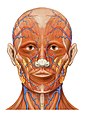
 The arteries of the face and scalp (angular artery labeled at center right) | |
| Details | |
| Source | Facial artery |
| Vein | Angular vein |
| Supplies | Lacrimal sac, orbicularis oculi muscle |
| Identifiers | |
| Latin | arteria angularis |
| TA98 | A12.2.05.029 |
| TA2 | 4397 |
| FMA | 49583 |
| Anatomical terminology | |
The angular artery is an artery of the face. It is the terminal part of the facial artery. It ascends to the medial angle of the eye's orbit. It is accompanied by the angular vein. It ends by anastomosing with the dorsal nasal branch of the ophthalmic artery. It supplies the lacrimal sac, the orbicularis oculi muscle, and the outer side of the nose.
Structure
The angular artery is the terminal part of the facial artery.[1][2] It ascends to the medial angle of the eye's orbit (the medial canthus).[2] It is embedded in the fibers of the angular head of the levator labii superioris muscle.[citation needed] It is accompanied by the angular vein. On the cheek, it distributes branches which anastomose with the infraorbital artery.[2] It ends by anastomosing with the dorsal nasal branch of the ophthalmic artery.
Function
The angular artery supplies the lacrimal sac,[2] most of the outer side of the nose,[3] part of the lower eyelid,[2] and the orbicularis oculi muscle.[citation needed]
Clinical significance
The angular artery is important in a nasolabial skin flap for reconstructive surgery.[4] It can be put at risk during acupuncture of skin around the inner side of the eye.[5]
Additional images
-
 Lateral head anatomy detail
 -
 Head anatomy anterior view


References
![]() This article incorporates text in the public domain from page 556 of the 20th edition of Gray's Anatomy (1918)
This article incorporates text in the public domain from page 556 of the 20th edition of Gray's Anatomy (1918)
- ^ Scremin, Oscar U. (2015). "31 - Cerebral Vascular System". The Rat Nervous System (4th ed.). Academic Press. pp. 985–1011. doi:10.1016/B978-0-12-374245-2.00031-0. ISBN 978-0-12-374245-2.
- ^ a b c d e Remington, Lee Ann (2012). "11 - Orbital Blood Supply". Clinical Anatomy and Physiology of the Visual System (3rd ed.). Butterworth-Heinemann. pp. 202–217. doi:10.1016/B978-1-4377-1926-0.10011-6. ISBN 978-1-4377-1926-0.
- ^ Ort, Yirae; Quereshy, Faisal A. (2017). "83 - Basic Rhinoplasty". Maxillofacial Surgery. Vol. 2 (3rd ed.). Churchill Livingstone. pp. 1257–1278. doi:10.1016/B978-0-7020-6056-4.00083-6. ISBN 978-0-7020-6056-4.
- ^ Markiewicz, Michael R.; Ord, Robert; Fernandes, Rui P. (2017). "43 - Local and Regional Flap Reconstruction of Maxillofacial Defects". Maxillofacial Surgery. Vol. 1. Churchill Livingstone. pp. 616–635. doi:10.1016/B978-0-7020-6056-4.00044-7. ISBN 978-0-7020-6056-4.
- ^ Focks, Claudia; März, Ulrich (2008). "4 - Acupuncture Points of the Twelve Primary Channels". Atlas of Acupuncture. Churchill Livingstone. pp. 223–241. doi:10.1016/B978-044310028-4.50007-2. ISBN 978-0-443-10028-4.
External links
- Articles with short description
- Short description matches Wikidata
- Anatomy NAV infobox with use of other NAV parameters
- All articles with unsourced statements
- Articles with unsourced statements from February 2022
- Wikipedia articles incorporating text from the 20th edition of Gray's Anatomy (1918)
- Articles with TA98 identifiers
- Arteries of the head and neck
- Human head and neck
- Otorhinolaryngology
- Cardiovascular system