File:PMC4362101 SaudiMedJ-35-S49-g007.png

From WikiProjectMed
Jump to navigation Jump to search

PMC4362101_SaudiMedJ-35-S49-g007.png(512 × 194 pixels, file size: 105 KB, MIME type: image/png)

This file is from a shared repository and may be used by other projects. The description on its file description page there is shown below.

Summary

Description
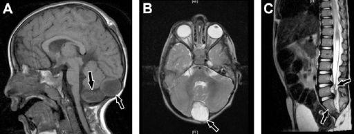
English: F6: Caudal regression syndrome. A) Brain MRI showing 2 cystic structures at the region of cisterna magna demonstrating high signal intensity on T1-weighted image (arrows). B) A cyst is also shown on axial T2-weighted image (arrow). C) Sagittal MRI dorsolumbar spine showing abrupt cutoff (truncation) and bulbar configuration of conus medullaris at the level of L1 vertebral body and thickening of filum terminale (right arrow). There is also sacral agenesis (only 2 sacral segments are visualized, left arrow).
Date
Source https://openi.nlm.nih.gov/detailedresult?img=PMC4362101_SaudiMedJ-35-S49-g007&query=Caudal%20regression%20syndrome&it=xg&req=4&npos=2
Author Seidahmed MZ, Abdelbasit OB, Shaheed MM, Alhussein KA, Miqdad AM, Samadi AS, Khalil MI, Al-Mardawi E, Salih MA

Licensing

English: This file is licensed CC BY-NC-SA 3.0

This file was uploaded with UploadWizard.

File history

Click on a date/time to view the file as it appeared at that time.

Date/TimeThumbnailDimensionsUserComment
current21:56, 21 June 2022Thumbnail for version as of 21:56, 21 June 2022512 × 194 (105 KB)Ozzie10aaaaUploaded a work by Seidahmed MZ, Abdelbasit OB, Shaheed MM, Alhussein KA, Miqdad AM, Samadi AS, Khalil MI, Al-Mardawi E, Salih MA from https://openi.nlm.nih.gov/detailedresult?img=PMC4362101_SaudiMedJ-35-S49-g007&query=Caudal%20regression%20syndrome&it=xg&req=4&npos=2 with UploadWizard

There are no pages that use this file.