File:PMC3181860 DialoguesClinNeurosci-10-47-g003.png
Jump to navigation
Jump to search
No higher resolution available.
PMC3181860_DialoguesClinNeurosci-10-47-g003.png (512 × 354 pixels, file size: 129 KB, MIME type: image/png)
File history
Click on a date/time to view the file as it appeared at that time.
| Date/Time | Thumbnail | Dimensions | User | Comment | |
|---|---|---|---|---|---|
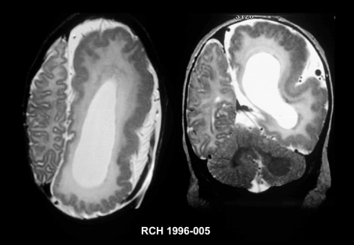
| current | 20:13, 10 January 2022 | | 512 × 354 (129 KB) | Ozzie10aaaa | Author:Leventer RJ, Guerrini R, Dobyns WB,Children's Neuroscience Centre & Murdoch Children's Research Institute, Royal Children's Hospital(Openi/National Library of Medicine) Source:https://openi.nlm.nih.gov/detailedresult?img=PMC3181860_DialoguesClinNeurosci-10-47-g003&query=Hemimegalencephaly&it=xg&req=4&npos=1 Description:DialoguesClinNeurosci-10-47-g003: Imaging features of hemimegalencephaly. Axial T2-wieghted MRI (left) and coronal T2 -weighted MRI (right) of an infant with hemimegalen... |
File usage
The following page uses this file: