File:CSF circulation.png
Jump to navigation
Jump to search
Size of this preview: 800 × 350 pixels. Other resolutions: 320 × 140 pixels | 640 × 280 pixels | 1,024 × 448 pixels | 1,956 × 856 pixels.
Original file (1,956 × 856 pixels, file size: 673 KB, MIME type: image/png)
File history
Click on a date/time to view the file as it appeared at that time.
| Date/Time | Thumbnail | Dimensions | User | Comment | |
|---|---|---|---|---|---|
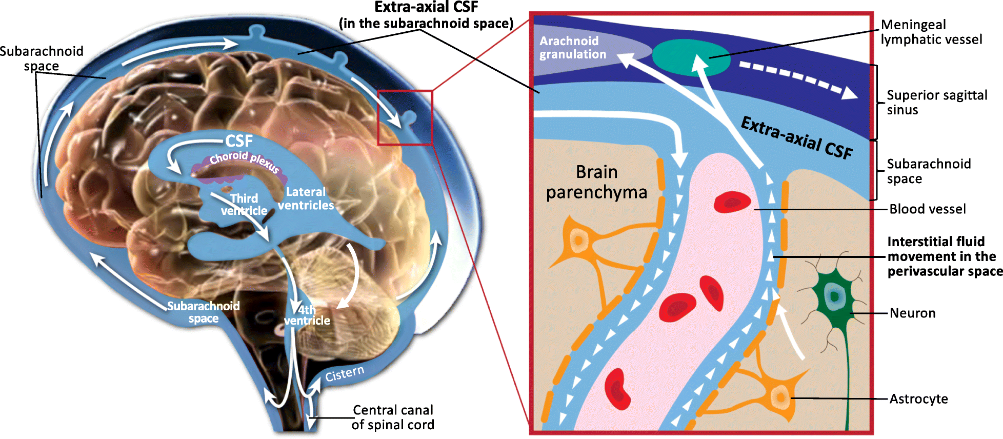
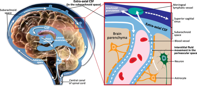
| current | 11:07, 16 June 2019 | | 1,956 × 856 (673 KB) | commons>Was a bee | {{Information |Description={{en|1=Schematic of CSF circulation, CSF outflow systems, and the anatomy of various CSF compartments. CSF is produced by the choroid plexus in the ventricles, where it delivers growth factors to progenitor cells that originate on the surface of the ventricles, and then proliferate into neurons and migrate to form the cerebral cortex. CSF circulates from the lateral, third and fourth ventricles to the cisterns of the brain, and then flows into the subarachnoid space... |
File usage
There are no pages that use this file.